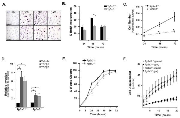
Figure 2. Cultured Tgfbr3−/− epicardial cells show decreased proliferation and invasion.
(A) Photomicrographs of cells incubated with BrdU and fixed at 24, 48 and 72h after initial seeding on 4-well collagen coated slides. (B) Quantitation of percent BrdU incorporation (n=3,*p=0.001) (C) Measurement of cell number by MTS assay (experiments were repeated 3 times in triplicate, results for one littermate pair shown, *p<0.05). (D) Quantitation of invasion using a modified Boyden chamber assay of one littermate pair (experiments were repeated 3 times in replicates of 6,*=p<0.05). (E) Graph quantifying percent wound closure over 72 hours after confluent cell monolayers were wounded using a rotating silicon tip (experiments were repeated three times, results for one littermate pair shown, *p<0.05). (F) Mean cell displacements values using live video microscopy of one littermate pair. The difference between genotypes is not significant.

