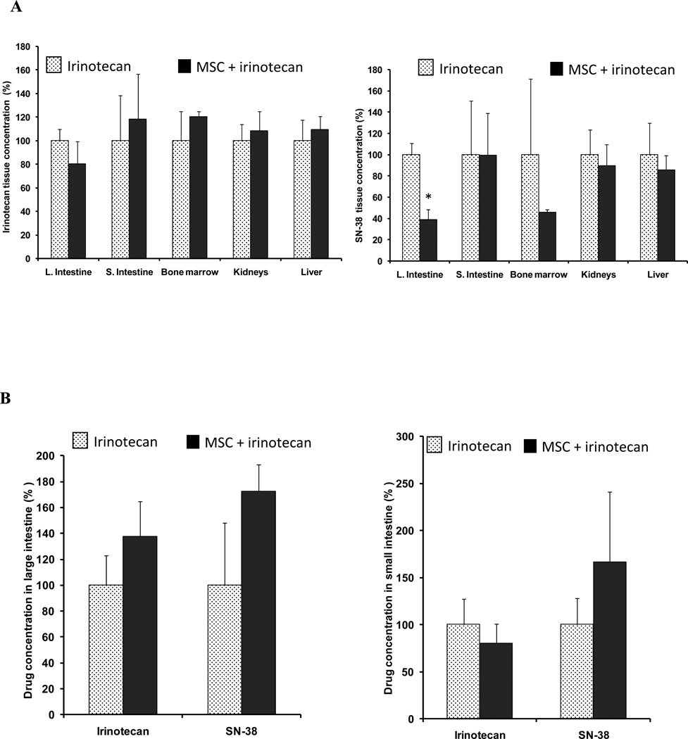
Figure 4. Drug concentrations in normal tissue.
Panel A shows irinotecan or SN-38 concentrations in large intestine, small intestine, bone marrow, kidneys and liver 2h after treatment with 100mg/kg irinotecan alone  , or concurrent combination treatment of MSC (0.2 mg/d) and irinotecan
  .
.
Panel B shows irinotecan and SN-38 concentrations in large intestine or small intestine 2h after the second course of 100mg/kg irinotecan alone  , or in combination with MSC
  .
.
When compared with irinotecan alone, the only significant change in drug concentration was a decrease in large intestine SN-38 concentration in panel A (p<0.05). * denotes p < 0.05 when compared to irinotecan alone.

