Figure 5.
Induction of intra-tumor microvessel maturation after the second course of the sequential combination treatment
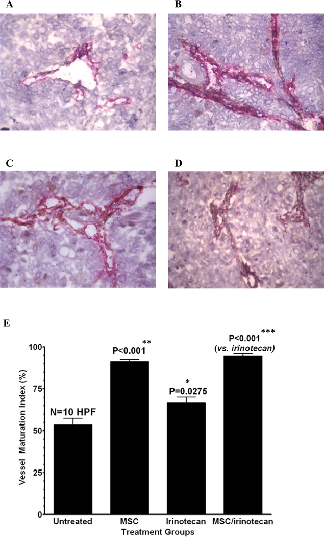
MSC enhances intra-tumor microvessel maturation as studied on CD31/α-SMA double stained (CD31 for endothelial cells: red, α-SMA for pericytes: brown) frozen sections of FaDu xenografts on day 15 after various treatments (Panel A: untreated, B: MSC, C: irinotecan, D: irinotecan/MSC). The bar graphs show the corresponding vascular maturation index (VMI) which is generally used as a quantitative measure for vascular maturation. The photomicrographs visualize the trend of the change in the pattern of double-immunostaining of the vessels, illustrating that the brown component for pericytes is increasing if MSC treatment is involved (panel B versus A and panel D versus C).
The date of relevant bar graphs from computer image analysis numerically demonstrates that MSC alone and in combination significantly enhanced the VMI as compared to the control or CPT induced VMI, respectively. It proves that MSC treatment increases the proportion of pericytes associated with endothelial cells which is an indication of vessel maturation. CPT treatment alone also increased VMI, but less effective than MSC alone, as compared to the control. * denotes p < 0.05 when compared to untreated controls, ** denotes p < 0.001 when compared to irinotecan alone and *** denotes p < 0.001 when compared to untreated controls.

