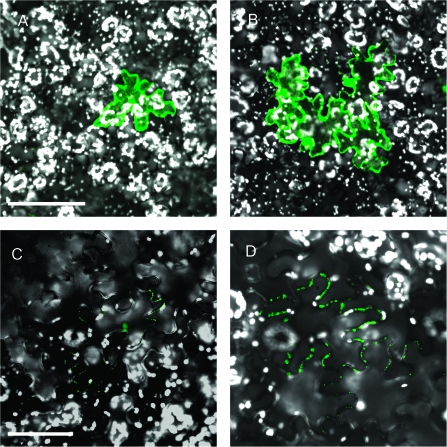
Figure 1.
Cell-to-Cell Transport of Viral MPs.
Constructs encoding CaLCuV MP-GFP (A, B) or TMV MP–YFP (C, D) were transiently expressed in N. benthamiana epidermal cells by biolistic bombardment (Ueki et al., 2009, 2010a). In some cases, the expressed proteins remained confined in single cells (A, C), indicating the lack of movement. In other cases, the proteins moved from cell to cell, resulting in signal clusters composed of several cells (B, D). These differences in the movement capacity most likely reflect the host mechanisms that restrict viral movement as described in the text. Bars are 100 and 50μm in panels (A) and (C), respectively. All images represent single confocal sections.

