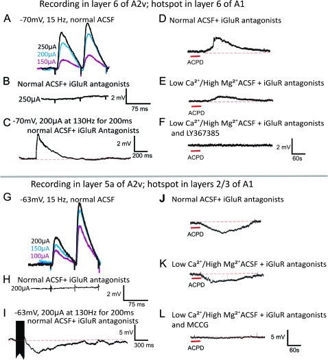
Figure 4.
Postsynaptic activation of mGluRs in the A1 to A2v pathway. (A–F) Example showing activation of mGluR1s with paired-pulse facilitation (A), responses to low-frequency stimulation blocked by iGluR antagonists (B), and evidence of mGluR activation (C). With normal ACSF and iGluR antagonists, the addition of ACPD evokes a sustained depolarization (D), which is also seen after a switch to high-Mg2+/low-Ca2+ ACSF and iGluR antagonists (E). Subsequent addition of mGluR1 antagonist abolishes this response to ACPD (F). (G–L) Example in another neuron showing activation of Group II mGluRs; conventions as in A–F, but in this case the activation of the mGluRs is hyperpolarizing.

