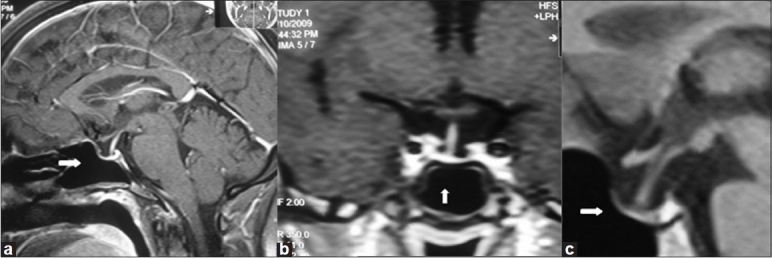
Figure 1.

MRI pituitary (a) Sagital view case 1, (b) Coronal view case 3, (c) Sagital view case 4 showing pituitary fossa filled with cerebrospinal fluid and stalk touching the base of pituitary floor (white arrows); features suggestive of empty sella
