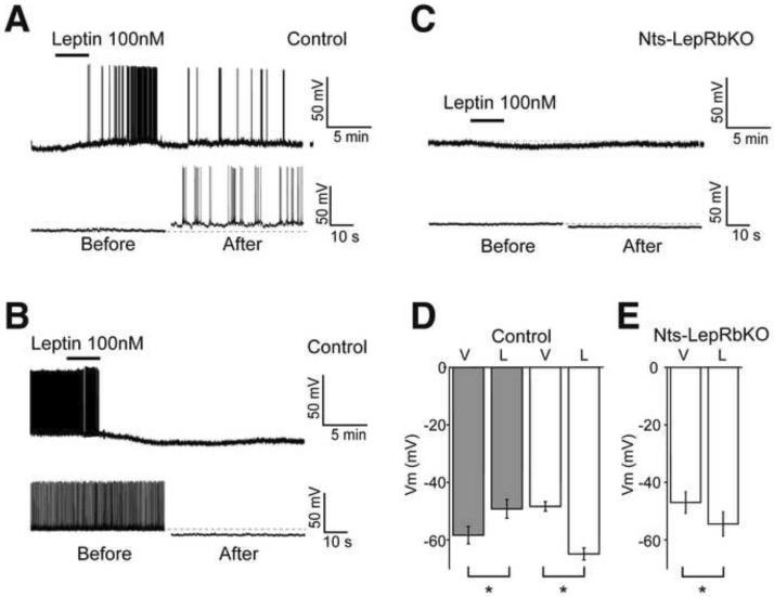
Figure 2. Electrophysiology of LHA Nts Neurons in Response to Leptin.
Brain slice recordings of EGFP (i.e., Nts) neurons in the LHA of Nts-EGFP (Control) and Nts-LepRbKO-EGFP (Nts-LepRbKO) mice treated with vehicle (V) or leptin (100 nM, L). Representative traces (A, B, C; bottom panel, on the expanded time scale) and aggregate responses (D, E) from leptin-depolarized (A, D-grey bars) and leptin-hyperpolarized neurons (B, D- white bars) in Control mice, and (C, E) all recorded neurons in Nts-LepRbKO mice. All the recordings were performed in the presence of GABAA, glycine and glutamate receptor antagonists. Data represent mean ± SEM, * = p<0.01 by paired t test.

