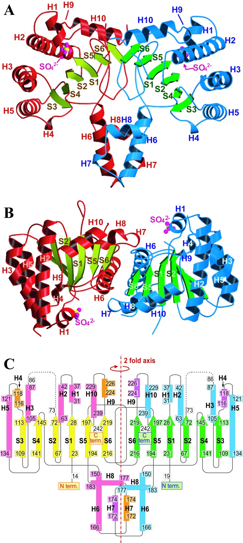
Figure 2.
Overall structure of UPS from M. luteus B-P 26. A front view (A) and a top view (B) of the dimer structure (ribbon model). One monomer of UPS is shown by red helices (α- and 310-helices) and yellowish green arrows (β-strands), and the other monomer by blue helices and green arrows. Helices (H1–H10) and strands (S1–S6) are labeled together with the sulfate ions found in the crystal structure. These two figures were prepared with molscript (33) and raster3d (34). (C) A topology diagram of the secondary structure of the UPS dimer. α- and 310-helices of one monomer and those of the other monomer are colored pink, orange, sky-blue, and light purple, respectively. β-strands of one monomer and the other are colored yellow and green, respectively.

