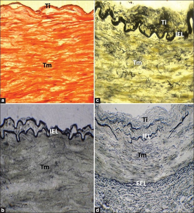
Figure 1.

(a) RA of a 25-year-old individual, stained with H and E, showing no intimal changes (×400). (b) The cross section of RA of a 20-year-old individual, stained with VVG stain, showing duplicated IEL (×400). (c) RA of a 45-year-old individual, stained with VVG stain, showing intimal thickening and numerous fine elastic fibers in Tm (×400). (d) Disorganized IEL with elastic fibers in Tm in RA of a 59-year-old individual, stained with VVG stain. EEL was well defined and appears intact in the entire periphery of the vessel wall (×200). Ti: Tunica intima; Tm: Tunica media; IEL: Internal elastic lamina; EEL: External elastic lamina
