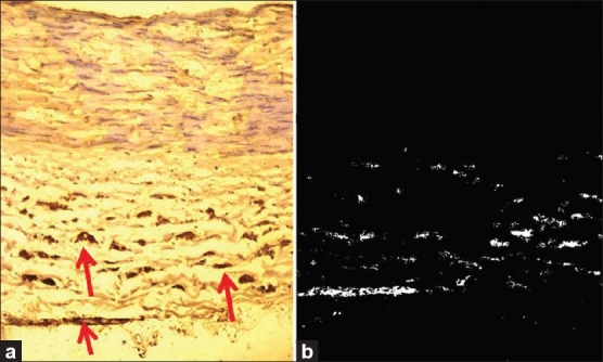
Figure 2.

(a) Arrows pointing to the sympathetic fibers in a RA of a 26-year-old individual, stained with TH immunostaining (×250). (b) Results of the automated measurement of sympathetic fiber area (white dots) of the same RA that was calculated by Tissue Quant image analysis software (×250)
