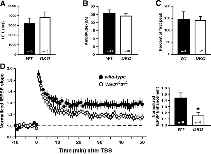
Figure 5.
Vav GEFs mediate synaptic plasticity in the hippocampus. A, Average IEI per neuron from wild-type or Vav2−/−3−/− CA1 pyramidal neurons (n.s., Student's t test). B, Average mEPSC amplitude per neuron from same recordings as in B (n.s., Student's t test). C, Paired-pulse stimulation (10 Hz). Average paired-pulse ratio measured as the percentage of the second pulse relative to the first (n.s., Student's t test). D, Left, fEPSPs were recorded from CA1 of P15 wild-type or Vav2−/−3−/− acute hippocampal slices (300 μm) in response to two theta burst stimulations of Schaffer collaterals. Right, The fEPSP slopes were normalized for each genotype. Normalized fEPSP enhancement at 40–50 min is shown (*p < 0.05, Student's t test).

