
Figure 4.
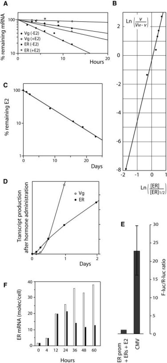
Quantification experiments. (A) Degradation rates of ER and Vg mRNA in cultured trout hepatocytes (circles, ER; diamonds, Vg; open symbols, without hormone; solid symbols, with 1 μM hormone). (B) Cooperative ER concentration-dependence of the Vg gene transcription rate (v). (C) Rate of clearance of estradiol in vivo in a male rainbow trout, determined by radio-immunoassay. (D) Transcript accumulation for Vg (open circles) and ER (solid circles), determined by simultaneous run-on analyses of the ER and Vg gene transcription rates in nuclei isolated from hormone-stimulated liver cells. (E) Comparative maximal expression in HeLa cells, of the CMV promoter and of the zebrafish ER gene promoter in presence of zERs and of 1 μM E2. HeLa cells were transfected with the short isoform of zebrafish ER and a F-Luc reporter gene driven by the zebrafish ER gene promoter. (F) Selective poisoning of the AF2/LBD domain using 4-OHT in cultured trout hepatocytes. Estradiol was added at time 0 and 4-OHT was added (solid histograms) or not (open histograms) 12 h later.