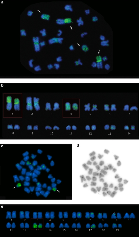
Figure 4.
(a) Cross-species fluorescence in situ hybridization (FISH) using Y-chromosome probe obtained from Eigenmannia sp.2 hybridized to an Eigenmannia sp.1 metaphase. Pairs displaying consistent signals on euchromatic regions are indicated. (b) Chromosomes from the same metaphase ordered by size to aid interpretation of signals. (c) Cross-species FISH using X-chromosome probe obtained from E. virescens (2n=38XX:XY) hybridized to an E. virescens metaphase. Pairs displaying consistent signals on euchromatic regions are indicated. (d) Inverted 4′-6-diamidino-2-phenylindole image from same metaphase (to facilitate visualization of chromosome morphology). (e) Chromosome pairs from panel c ordered by size.

