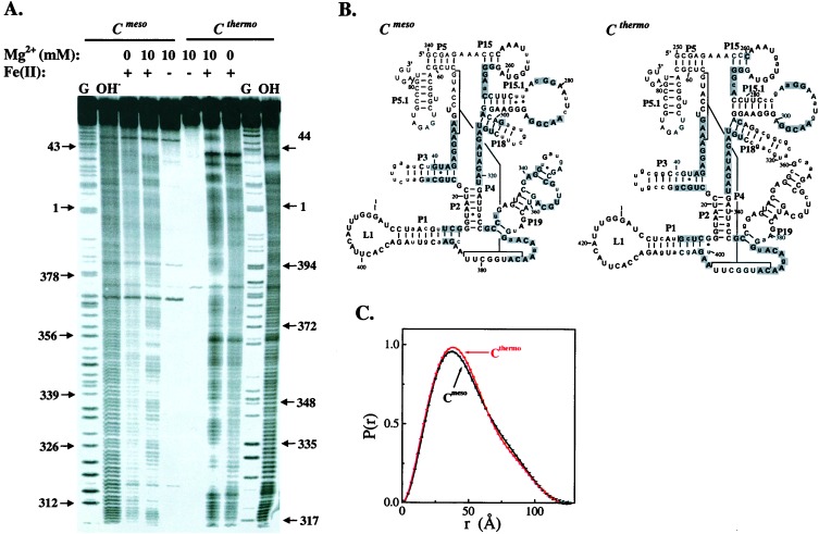
Figure 2.
Both ribozymes have similar native structures. (A) Hydroxyl radical protection in 20 mM Tris⋅HCl, pH 7.5, 10 mM MgCl2, 37°C (18, 26). (B) Protection mapped onto the phylogenetically derived secondary structure (35). Residues protected in the presence of Mg2+ are shaded. Differences in the sequence are shown in lowercase. Residues that cannot be analyzed are shown as smaller, plain letters. (C) Pair-distribution function of the native state determined by SAXS.

