Abstract
Cytoplasmic incompatibility (CI) allows the intracellular, maternally inherited bacterial symbiont Wolbachia to invade arthropod host populations by inducing infertility in crosses between infected males and uninfected females. The general pattern is consistent with a model of sperm modification, rescued only by egg cytoplasm infected with the same strain of symbiont. The predacious flower bug Orius strigicollis is superinfected with two strains of Wolbachia, wOus1 and wOus2. Typically, superinfections of CI Wolbachia are additive in their effects; superinfected males are incompatible with uninfected and singly infected females. In this study, we created an uninfected line, and lines singly infected with wOus1 and wOus2 by antibiotic treatment. Then, all possible crosses were conducted among the four lines. The results indicated that while wOus2 induces high levels of CI, wOus1 induces very weak or no CI, but can rescue CI caused by wOus2 to a limited extent. Levels of incompatibility in crosses with superinfected males did not show the expected pattern. In particular, superinfected males caused extremely weak CI when mated with either singly infected or uninfected females. An analysis of symbiont densities showed that wOus1 densities were significantly higher than wOus2 densities in superinfected males, and wOus2 densities were lower, but not significantly, in superinfected relative to singly infected males. These data lend qualified support for the hypothesis that wOus1 interferes with the ability of wOus2 to cause CI by suppressing wOus2 densities. To our knowledge, this is the first clear case of non-additive CI in a natural superinfection.
Keywords: reproductive parasites, unidirectional cytoplasmic incompatibility, bidirectional cytoplasmic incompatibility, multiple infections, predacious flower bug
Introduction
Wolbachia are maternally transmitted intracellular bacteria infecting a large proportion of arthropods and nematodes (Bandi et al., 1998; Stouthamer et al., 1999; Werren et al., 2008). A recent meta-analysis of Wolbachia surveys estimated that 66% of insect species harbor Wolbachia at varying frequencies (Hilgenboecker et al., 2008). Wolbachia infections can induce reproductive alterations such as feminization (Rigaud et al., 1997), thelytokous parthenogenesis (Stouthamer, 1997), male killing (Hurst et al., 2000) and cytoplasmic incompatibility (CI; Hoffmann and Turelli, 1997; Charlat et al., 2002) as well as influence the general fitness and life history of hosts in both positive and negative ways (for example Min and Benzer, 1997; Dobson et al., 2004). CI is the most common effect of Wolbachia and has been described in crustaceans (Moret et al., 2001), arachnids (Breeuwer, 1997) and in most groups of insects that have been examined (for reviews, see Stouthamer et al., 1999; Werren et al., 2008). The incompatibility occurs in crosses between males and females with differing Wolbachia infection status. It can be either unidirectional, when infected males mate with uninfected females, or bidirectional, in crosses in which both males and females are infected with different CI-inducing Wolbachia strains (Hoffmann and Turelli, 1997). Unidirectional CI can also occur between a CI-inducing strain and a strain that does not induce CI, where the incompatible crosses occur between males with the CI-causing strain and females of the non-CI strain, or uninfected females (Duron et al., 2006; Zabalou et al., 2008). Although the strain-specific mechanism of CI is still not well understood, the general pattern is consistent with a model in which sperm is modified in such a way that only the same strain of symbionts in the egg cytoplasm can rescue them and allow normal embryonic development (Werren, 1997). CI symbionts spread because infected females gain in fitness relative to uninfected females that can successfully reproduce only with uninfected males. Superinfections (that is co-infection with two or more Wolbachia strains) occur naturally (Sinkins et al., 1995; Zhou et al., 1998; Dobson et al., 2004) and have been generated artificially as well (Rousset et al., 1999; Walker et al., 2009). These superinfections typically have additive effects, such that a superinfected male is unidirectionally incompatible with both singly infected and uninfected females (Table 1), whereas superinfected females can rescue infections in both superinfected and singly infected females.
Table 1. Predicted results of crosses among hosts of two strains of Wolbachia (A and B) when the Wolbachia strains are both present in superinfected (A+B) individuals as well as in singly infected (A and B) individuals or are absent (U, uninfected).
| Female type |
Male type |
|||
|---|---|---|---|---|
| U | A | B | A + B | |
| U | + | − | − | − |
| A | + | + | − | − |
| B | + | − | + | − |
| A + B | + | + | + | + |
The pattern shown here pertains when both strains cause CI individually, and there is bidirectional incompatibility between the two strains. In general, incompatibility (−) is predicted when the male host harbors a Wolbachia strain not present in the female mate. Successful matings (+) occur when males have the same, or fewer strains of Wolbachia than the female. Note that superinfected males are predicted to be incompatible with all but superinfected females. (modified from Dobson et al., 2004).
The predacious flower bug Orius strigicollis (Poppoius) (Heteroptera: Anthocoridae) is one of the most useful biological control agents of various minute insect pests, such as thrips, and is common in agricultural fields in Japan. The species is also commercially available in Japan as a biological control agent for greenhouses (Yano, 2004). In this study, we show that this population of O. strigicollis is superinfected with Wolbachia, and the superinfection does not cause strong CI, even though one of the strains it carries causes strong CI when alone; this is not the expected additive effect of a superinfection (see Table 1). We speculate that the superinfection may have spread instead because of its superior ability to rescue CI.
Materials and methods
Insects
O. strigicollis was obtained from Sumitomo Chemical Co. Ltd. The strains were originated from Wakayama, Japan. O. strigicollis individuals were maintained in the laboratory (16 h light, 60% humidity, 25 °C) and reared on diet, Ephestia kuehniella Zeller (Lepidoptera: Pyralidae) eggs and pure water with cotton.
Wolbachia strains and origin of Orius lines of different infection status
The O. strigicollis culture we acquired was fixed for a superinfection of two different Wolbachia strains: wOus1 and wOus2 (wOus1+2). We note that natural populations of this species in different areas of Japan show more variation with respect to Wolbachia infection. In most populations, the greatest number of individuals is superinfected, but some singly infected individuals of both types are also found (Watanabe et al., submitted). The range of infection frequencies for wOus1+2, wOus1 and wOus2 in several populations are 23–100, 0–25 and 0–77%, respectively.
In this study, we used primers of the wsp region of Wolbachia for diagnostic PCR (see below). We previously examined another suite of Wolbachia genes, that is the housekeeping genes for MLST analysis, and also found consistent differences between the two Wolbachia strains in these genes (Baldo et al., 2006).
An uninfected line and lines singly infected with either wOus1 or wOus2 lines were derived from superinfection with wOus1+2. To establish these lines, superinfected wOus1+2 Orius were cured of one or more symbionts by feeding last-instar nymphs honey solution containing 50 mg ml−1 of tetracycline for 48 h in each of three consecutive generations. Nymphs treated with antibiotics were isolated until adults in each generation. In establishment of uninfected line, females mated with males of the same line. In establishment of wOus1 or wOus2 line, we used uninfected males to avoid incompatibility. After oviposition, we tested for infection status of females by diagnostic PCR. All crossing experiments were carried out at least five generations after the last antibiotic treatment to avoid any direct effect of the antibiotics.
DNA extraction and diagnostic PCR
To determine infection status of individuals of O. strigicollis, DNA was extracted in 32 μl STE buffer (5 NaCl, 500m EDTA (pH 8.0) and 1 Tris-HCl (pH 8.0)) and incubated with 2 μl of proteinase K (0.5 mg ml–1) at 56 °C for 2 h. The homogenate was heated at 99.9 °C for 5 min to inactivate the proteinase K, and then used as a template for PCR. PCR amplifications were conducted under the following conditions: 16.5 μl of 2 × AmpliTaq Gold PCR Master Mix (PE Applied Biosystems, Tokyo, Japan), 1.3 μl forward and reverse primer (10 pmol μl−1) and 13.9 μl of sterile water, a total of 33 μl PCR reaction volume. We monitored the infection status of the infected and uninfected laboratory stocks through PCR using the Wolbachia-specific wsp primers, wOus1-f (5′ TAA ATA CTT CTG AAA CAA ATG TTG 3′) and wOus1-r (5′ AAA AAT TAA ACG CTA CTC CA 3′), wOus2-f (5′ GAT GTA GTA TCT GAT GAC AAG 3′) and wOus2-r (5′ GGA CGT TGA TCT CTT TAG TAG 3′) (Watanabe et al., submitted). Each Wolbachia sequence was deposited in GenBank (wOus1: AB094361, wOus2: AB094365). PCR amplification was carried out in an ABI thermocycler (PE Applied Biosystems PCR System 9700, PE Applied Biosystems) with the following thermal profiles: 95 °C for 10 min; 35 cycles of 94 °C for 1 min, 55 °C for 1 min 30 s and 72 °C for 1 min 30 s, followed by incubation at 72 °C for 1 min 30 s. The PCR included a negative control (sterile water instead of DNA) to detect contamination. The PCR products were resolved on a 1.5% agarose gel, stained with ethidium bromide and visualized under an UV transilluminator. To confirm that DNA was properly extracted, ITS primers were used to amplify nuclear ribosomal DNA as a positive control for the template DNA quality (Hinomoto et al., 2004).
Crossing experiments
Crossing experiments were performed to reveal how the different Wolbachia strains influenced CI modification and rescue. Four-day-old virgin females, either uninfected or infected with wOus1+2, wOus1 or wOus2 were isolated and individually mated with wOus1+2, wOus1, wOus2 and uninfected males (also 4 days old). Each of the 16 crosses was replicated 7–12 times. All mating was observed, and subsequently each female was allowed to oviposit onto a plant, Sedum rubrotinctum with diet in a vial (1 cm diameter × 5 cm length). The plants that served as oviposition substrates were removed every second day, and replaced with a fresh plant, whereas the eggs laid on the older foliage were counted. Females were allowed to oviposit for 8 days. Egg-hatching rates were scored 1 week after egg collection. Parents of each cross were tested by PCR for confirmation of the expected Wolbachia infection status. Egg-hatching rates were compared among the treatments by means of a logistic regression, a generalized linear model specially designed for modeling binomial data using the logistic-link function (McCullagh and Nelder, 1989; Wajnberg and Haccou, 2008). Computations were performed using SAS software (SAS Institute Inc., 1999). The sex ratio was tested by the Fisher's exact test using R ver. 2.7.1 software (R Development Core Team, 2005).
Comparative fecundity of uninfected, singly infected and superinfected lines
To determine whether female Orius with different symbiont combinations differ in fecundity, 4-day-old females of all four types were mated with 4-day-old males of the same strain for 48 h in plastic vials (1 cm diameter × 5 cm length). Then, females were placed in another vial of the same type as mentioned above and supplied with a plant for oviposition so that host fecundity could be evaluated. We counted the number of eggs at 2, 4, 6 and 8 days. The total number of eggs in each treatment produced by superinfected, uninfected and singly infected females of both types was analyzed with a repeated measures generalized linear model specially designed for modeling count data using a log-link function (McCullagh and Nelder, 1989; Wajnberg and Haccou, 2008). Computations were performed using SAS software (SAS Institute Inc., 1999).
Quantitative PCR
We performed quantitative PCR analysis of host abdomens. These tissues were isolated from individuals from superinfected and singly infected lines. Individual insects were carefully dissected with fine forceps under a binocular microscope. Isolated tissues were immediately subjected to DNA extraction. Quantitative PCR was carried out with the ABI PRISM 7000 Sequence Detection System (PE Applied Biosystems). Reaction volumes of 25 μl contained 12.5 μl of SYBR Green PCR Master Mix (PE Applied Biosystems), 10.5 μl of sterile water, 0.5 μl each of 10 μ forward and reverse primer, and 1 μl of target DNA in single wells of a 96-well plate (PE Applied Biosystems). For the selective amplification of a small portion of the O. strigicollis COI gene (90 bp) and Wolbachia wsp (wOus1, 85 bp and wOus2, 75 bp), the following primers were designed and used: OSCO1-F (5′-CTG CCC CCA TCT ATT ACA TTA CTT ATT-3′), OSCO1-R (5′-TGC TGA AAG AGG AGG ATA TAC TGT TC-3′), wOus1wsp-F (5′-ATG TTG AAG GGC TTT ACT CAC AAT T-3′), wOus1wsp-R (5′-GCT GTT AAA CTG TCT GCA ACA TTT G-3′), wOus2wsp-F (5′-TCA CAA TTG ACT AAA GAT GCA ACT GT-3′), wOus2wsp-R (5′-CAA TCC TGA AAA CGC TGT TAC ACT-3′). PCR primers and probes were designed using PRIMER EXPRESS 1.5 (PE Applied Biosystems). Cycle parameters were 50 °C for 2 min and 95 °C for 10 min to activate AmpliTaq Gold for preventing primer dimers, followed by 40 cycles of 95 °C for 15 s and 60 °C for 1 min. The number of each Wolbachia and COI genes were calculated from the intensity of the fluorescence on the basis of a standard curve obtained from standard samples.
To compare Wolbachia density between superinfected and singly infected abdomens, we analyzed the data using a Student's t-test (R software ver. 2.7.1; R Development Core Team, 2005).
Results
CI modification
Our results suggested that the two Wolbachia strains differed in their ability to cause CI. In the predicted incompatible cross with wOus1 (wOus1 males × uninfected females), the mean egg-hatch rate was 81.0%, not significantly different from either of the predicted control crosses (the reciprocal cross), Table 2 (χ2=0.03, df=1, P=0.9) and wOus1 males × females infected with the same strain (χ2=0.06, df=1, P=0.8). On the other hand, the single infection of wOus2 induced high levels of CI in the predicted CI cross (wOus2 males × uninfected females), resulting in approximately 10% of the hatching rates of the control crosses. In crosses between uninfected females and singly infected wOus2 males, only 35 of 388 eggs hatched (Table 2). This egg-hatching rate is significantly lower than for the reciprocal cross (χ2=173.30, df=1, P<0.0001), or for the within-strain cross (χ2=168.37, df=1, P<0.0001).
Table 2. Egg-hatch rate (mean % ± s.d.) of progeny of crosses among lines of Orius strigicollis that are superinfected (wOus1 and 2), singly infected (wOus1 and 2), and uninfected with Wolbachia.
| Female type |
Male type |
||||
|---|---|---|---|---|---|
| Uninfected | wOus1 | wOus2 | wOus1+2 | ||
| Uninfected | 83.7±7.1abc | 81.0±6.4bcd | 8.5±8.6g | 56.4±18.2e | 58.6±31.6A |
| (12, 415) | (10, 370) | (10, 388) | (12, 385) | (44, 1558) | |
| wOus1 | 82.9±9.2bcd | 82.4±4.2bcd | 20.5±14.9f | 49.6±18.2e | 61.1±27.7A |
| (12, 449) | (12, 463) | (9, 248) | (13, 410) | (46, 1570) | |
| wOus2 | 89.4±5.3a | 73.5±7.6d | 85.7±7.1ab | 76.4±9.3cd | 81.6±9.7B |
| (10, 368) | (7, 214) | (10, 385) | (12, 635) | (39, 1602) | |
| wOus1+2 | 85.7±7.6ab | 81.0±7.7bcd | 86.6±7.1ab | 84.7±4.0ab | 84.8±6.7B |
| (10, 514) | (7, 311) | (10, 361) | (10, 411) | (37, 1597) | |
| 85.2±7.7A | 80.0±6.9B | 51.1±37.9C | 65.7±19.8D | ||
| (44, 1746) | (36, 1358) | (39, 1382) | (47, 1841) | ||
Numbers within parentheses refer to number of pairs (n) and total number of eggs counted for each cross.
The far right column provides means of all crosses by particular female types, and the bottom row provides means of all crosses by particular male types. Mean frequencies marked with the same lower-case letter (in cells) or upper-case letter (in marginal rows or columns) are not significantly different (logistic regression, P>0.05).
Surprisingly, given the strong CI induced by wOus2, superinfected males caused only weak CI when mated with uninfected females. The egg-hatch rate of this cross was, on average, about two-thirds that of compatible crosses, and quite variable. The egg-hatch rate for this cross was statistically significantly above that for the wOus2 incompatible cross (χ2=86.59, df=1, P<0.0001), but also significantly below the rate of egg hatch of the control reciprocal cross (χ2=44.52, df=1, P<0.0001), and the within-strain cross (χ2=33.91, df=1, P<0.0001).
CI rescue
The Wolbachia strains also appeared to vary in their ability to rescue CI. Although wOus1 caused either weak or no CI, it showed some ability to rescue sperm modified by wOus2; the egg-hatch rates of wOus1 females mated to wOus2 males were double that of uninfected females × wOus2 males (χ2=106.14, df=1, P<0.0001). The rescue function of wOus1 females when mated to wOus2 or superinfected males was also variable. The apparent lack of rescue by wOus2 females was somewhat puzzling. The egg-hatch rates in the cross between wOus2 females and wOus1 males were significantly lower than when wOus2 females mated with males of the same type (χ2=33.91, df=1, P=0.0046). Similarly, hatch rates were lower when wOus2 females mated with superinfected males relative to within-superinfection matings (χ2=4.98, df=1, P=0.0256). The result is puzzling because, as discussed above, the degree of CI caused by wOus1 is very weak if it occurs at all; the egg-hatch rate in the predicted CI cross is not significantly different from the control crosses.
As expected, superinfected females rescued sperm from all male types. There were no significant differences among crosses between superinfected females and uninfected, singly infected or superinfected males (Table 2).
Comparative fitness assays
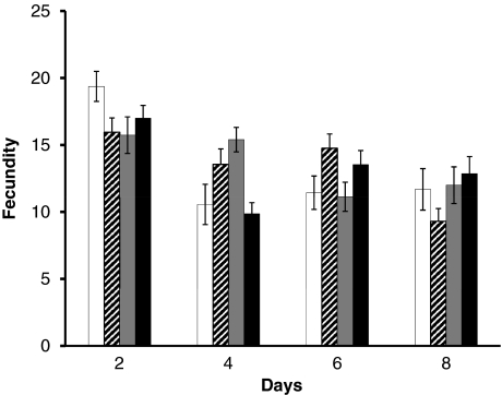
In all crosses, the sex ratio of O. strigicollis developed to adults showed no significant differences from 1:1 (Fisher's exact test, P>0.05). There were also no significant differences between the number of eggs produced by singly infected, superinfected or uninfected females on each date (Figure 1).
Figure 1.
Fecundity of superinfected, singly infected and uninfected females. Data are shown as mean ± s.e. of offspring produced by females at days 2, 4, 6 and 8. There was no significant difference among singly infected (wOus1 or wOus2), superinfected (wOus1+2) and uninfected lines (repeated measures generalized linear model: χ2=0.22, df=3, P>0.05), although there was a significant ‘day' effect (χ2=29.61, df=3, P<0.001) and the ‘day' effect differs significantly among singly infected, superinfected and uninfected lines (interaction: χ2=36.66, df=9, P<0.001). White bars: uninfected (number of pairs, n=16); striped bars: wOus1 (n=20); gray bars (n=15): wOus2; black bars: wOus1+2 (n=15).
Infection densities of wOus1 and wOus2 in superinfected and singly infected strains
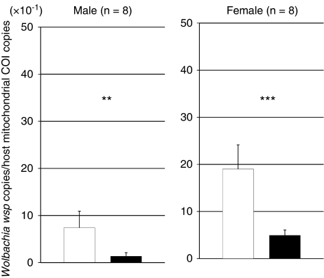
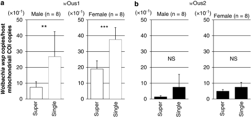
The infection densities of wOus1 Wolbachia in the abdomens of superinfected strains were much greater than those of wOus2 in both males and females (Figure 2; Student's t-test, male: P<0.01, female: P<0.001). Similarly, we examined the density of wOus1 and wOus2 in abdomens of singly infected individuals and compared them with the density of these Wolbachia strains in superinfected individuals. We found that the densities of wOus1 in superinfected individuals were significantly lower than in singly infected individuals (Figure 3a; Student's t-test, male: P<0.01, female: P<0.001). The densities of wOus2 were also lower in superinfected individuals than in singly infected individuals, but the data were more variable and the difference was not statistically significant (Figure 3b; Student's t-test, male: P=0.05127, female: P<0.001). These patterns were observed in both sexes.
Figure 2.
Relative infection densities of wOus1 and wOus2 in the abdomens of superinfected individuals. White bars: wOus1; black bars: wOus2. Different letters indicate statistically significant differences (Student's t-test, **P<0.01, ***P<0.001). Males and females were subjected to the statistical analysis separately.
Figure 3.
Relative infection densities of the two Wolbachia strains in the abdomens of singly vs superinfected individuals. White bars: wOus1 (a); black bars: wOus2 (b). Different letters indicate statistically significant differences (Student's t-test, NS P>0.05, ** P<0.01, *** P<0.001). Males and females were subjected to the statistical analysis separately.
Discussion
Here, we show that two strains of Wolbachia, one of which causes strong CI when alone, induce only weak CI in the superinfected state in the predacious bug O. strigicollis. The great majority of studies in which the compatibilities of superinfected, singly infected and uninfected lines have been studied show an additive effect; superinfected males cause CI similar to the strength of the strongest single infection when mated to singly or uninfected females (Sinkins et al., 1995; Perrot-Minnot et al., 1996; Mercot and Poinsot, 1998; Rousset et al., 1999; Dobson et al., 2001; Mouton et al., 2005). We are aware of only one other similar example from the nature of a lack of additive CI effects of superinfection, in field populations of spider mites infected with Cardinium (which causes CI when alone), Wolbachia (which does not) or both (with no CI induced) (Ros and Breeuwer, 2009). In the mite study, it was not feasible to manipulate the infections of mites, however, and, therefore, not possible to determine whether the different phenotypes were due to strain differences in the Cardinium in the single and superinfected populations, or interference between the symbionts in the superinfected mites. In another, even more pertinent example, Walker et al. (2009) created artificial superinfections of mosquitoes by adding a Wolbachia strain that induced CI to a host bearing one that did not. In the superinfected lines, the new strain was stably maintained, but CI was not induced. Further, CI induced by the singly infected males was not rescued by superinfected females, likely because the density of the introduced symbiont was very low in both males and females (Walker et al., 2009). To our knowledge, this study is unique in showing differences in phenotype between single and naturally superinfected lines in which the symbiont strains are identical in both types of infection. The superinfection of CI symbionts in O. strigicollis is not additive in its phenotype, and suggests interference between the symbionts such that the CI that is caused by wOus2 when alone is suppressed when it is in a host co-infected with wOus1. Interestingly, although the modification aspect of CI appears to be reduced in the superinfected males, superinfected females are fully capable of rescuing sperm from the strong CI-inducing strain, wOus2, distinct from Ros and Breeuwer (2009) and Walker et al. (2009) examples, in which the rescue function of superinfected females was also impaired.
How would a superinfection that causes only weak CI spread in a population with a strong CI-inducing strain? Theory predicts that the spread and maintenance of a strictly maternally inherited symbiont is dependent on infected mothers producing more infected daughters than the number of daughters produced by uninfected females (Bull, 1983). This theory was recently extended to consider the evolution of symbiont densities in superinfected hosts (Engelstadter et al., 2007); superinfections are predicted to be stable only when superinfected females produce more infected daughters than singly or uninfected females (Engelstadter et al., 2007). We hypothesize that the first infection in O. strigicollis was wOus2. Theory would predict this symbiont would rapidly spread to fixation, given that CI is strong, and fecundity costs appear to be low or absent (Figure 1; Hoffmann and Turelli, 1997). Although the superinfection of wOus1 and wOus2 does not cause strong CI, it does induce weak CI and thus reduces the fitness of wOus2 females (Table 2). It also shows little or no cost to fecundity (Figure 2), rescues wOus2 sperm and appears to be marginally better at rescuing wOus1 sperm than wOus2 females as well, although this last difference is not statistically significant (Table 2). Given perfect co-transmission, and any slight superiority in its ability to rescue, we might predict that the superinfected females could produce more (superinfected) daughters than the number of daughters produced by wOus2 or uninfected females, and a superinfection would, therefore, invade.
The finding that superinfected males cause much weaker CI than does one of the strains while acting alone suggests some kind of interference of one strain with the other. Perhaps the most intuitive explanation would involve simple differences in density in the singly and superinfected hosts. At least in some hosts, CI intensity is related to symbiont density (Breeuwer and Werren, 1993; Hoffmann and Turelli, 1997). We predicted that, if density differences underlie the non-additive phenotype of superinfected hosts, then (1) the densities of wOus1 would be greater than wOus2 in doubly infected hosts. This is what we have observed (Figure 2), indicating the potential for competitive exclusion of wOus2 in tissues occupied by wOus1. This could be further tested by localizing the symbionts in tests with singly and doubly infected males. (2) We also predicted densities of wOus2 to be lower in superinfected males relative to singly infected males. The densities of wOus2 are indeed lower in doubly infected males, but the difference is not statistically significant, probably because the densities are quite variable (Figure 3b). Interestingly, there is also considerable variability in the egg-hatch rates when superinfected males mate with either uninfected or wOus1 females (Table 2), indicating that both wOus2 densities and CI intensity vary among individual superinfected males. In future work, it would be interesting to correlate the degree of CI and density of wOus2 and wOus1 in superinfected males to test the possibility that wOus2 densities and CI intensity co-vary. In summary, our results provide qualified support for the hypothesis that the non-additivity of CI expression in doubly infected males is mediated by a suppression of the density of wOus2. More work remains to be performed to resolve this question. However, suppression of the density of one symbiont by the other has been found before in natural infections (Kondo et al., 2005), and density differences may also have been the reason for the lack of CI modification and rescue in the Wolbachia superinfection created in mosquitoes by Walker et al. (2009). The higher densities of wOus1 compared with wOus2 suggest that wOus1 might also show a higher rate of maternal transmission in the field; this hypothesis should be investigated.
Lastly, the results presented here may temper our expectations about the potential applications of CI-inducing symbionts as tools in pest suppression when the pest/host population is already infected. Symbionts that cause CI have been considered as a means to directly reduce population size (Brelsfoard et al., 2008), limit longevity of disease vectors (McMeniman et al., 2009) and as a drive system for a beneficial gene (Beard et al., 1993; Bourtzis, 2008). When superinfections are not costly, and the phenotype of the superinfected individuals is the same level of CI as the single infection, there appears to be little cost to ‘stacking symbionts' in hosts (Sinkins et al., 1995). However, this example shows that adding symbionts that cause CI when alone may or may not result in the expected phenotype in a superinfected host.
Acknowledgments
We thank Dr Tetsuya Adachi-Hagimori and Eisuke Watanabe for helpful advice on the paper. We also thank three anonymous reviewers for helpful comments on previous versions of the paper.
The authors declare no conflict of interest.
References
- Baldo L, Hotopp J, Jolley K, Bordenstein SR, Biber S, Choudhury R, et al. Multilocus sequence typing system for the endosymbiont Wolbachia pipientis. Appl Environ Microbiol. 2006;72:7098–7110. doi: 10.1128/AEM.00731-06. [DOI] [PMC free article] [PubMed] [Google Scholar]
- Bandi C, Anderson TJ, Genchi C, Blaxter ML. Phylogeny of Wolbachia in filarial nematodes. Proc R Soc Lond B Biol Sci. 1998;265:2407–2413. doi: 10.1098/rspb.1998.0591. [DOI] [PMC free article] [PubMed] [Google Scholar]
- Beard CB, O'Neill SL, Tesh RB, Richards FF, Aksoy S. Modification of arthropod vector competence via symbiotic bacteria. Parasitol Today. 1993;9:179–183. doi: 10.1016/0169-4758(93)90142-3. [DOI] [PubMed] [Google Scholar]
- Bourtzis K.2008Wolbachia-based technologies for insect pest population controlIn: Aksoy S (eds).Transgenesis and the Management of Vector-Borne Disease Springer: New York; 104–113. [DOI] [PubMed] [Google Scholar]
- Breeuwer JAJ. Wolbachia and cytoplasmic incompatibility in the spider mites Tetranychus urticae and T. turkestani. Heredity. 1997;79:41–47. [Google Scholar]
- Breeuwer JAJ, Werren JH. Cytoplasmic incompatibility and bacterial density in Nasonia vitripennis. Genetics. 1993;135:565–574. doi: 10.1093/genetics/135.2.565. [DOI] [PMC free article] [PubMed] [Google Scholar]
- Brelsfoard CL, Sechan Y, Dobson SL. Interspecific hybridization yields strategy for South Pacific filariasis vector elimination. Plos Negl Trop Dis. 2008;2:e129. doi: 10.1371/journal.pntd.0000129. [DOI] [PMC free article] [PubMed] [Google Scholar]
- Bull JJ. Evolution of Sex Determining Mechanisms. Benjamin/Cummings Publ. Co.: Menlo Park, CA; 1983. [Google Scholar]
- Charlat S, Kirgianaki A, Bourtzis K, Mercot H. Evolution of Wolbachia-induced cytoplasmic incompatibility in Drosophilasimulans and D. echellia. Evolution. 2002;56:1735–1742. doi: 10.1111/j.0014-3820.2002.tb00187.x. [DOI] [PubMed] [Google Scholar]
- Dobson SL, Marsland EJ, Rattanadechakul W. Wolbachia-induced cytoplasmic incompatibility in single-and superinfected Aedes albopictus (Diptera: Culicidae) J Med Ent. 2001;38:382–387. doi: 10.1603/0022-2585-38.3.382. [DOI] [PubMed] [Google Scholar]
- Dobson SL, Rattanadechakul W, Marsland EJ. Fitness advantage and cytoplasmic incompatibility in Wolbachia single- and superinfected Aedes albopictus. Heredity. 2004;93:135–142. doi: 10.1038/sj.hdy.6800458. [DOI] [PubMed] [Google Scholar]
- Duron O, Bernard C, Unal S, Berthomieu A, Berticat C, Weill M. Tracking factors modulating cytoplasmic incompatibilities in the mosquito Culex pipiens. Mol Ecol. 2006;15:3061–3071. doi: 10.1111/j.1365-294X.2006.02996.x. [DOI] [PubMed] [Google Scholar]
- Engelstadter J, Hammerstein P, Hurst GDD. The evolution of endosymbiont density in doubly infected host species. J Evol Biol. 2007;20:685–695. doi: 10.1111/j.1420-9101.2006.01257.x. [DOI] [PubMed] [Google Scholar]
- Hilgenboecker K, Hammerstein P, Schlattmann P, Telschow A, Werren JH. How many species are infected with Wolbachia?–a statistical analysis of current data. FEMS Microbiol Lett. 2008;281:215–220. doi: 10.1111/j.1574-6968.2008.01110.x. [DOI] [PMC free article] [PubMed] [Google Scholar]
- Hinomoto N, Muraji M, Noda T, Shimizu T, Kawasaki K. Identification of five Orius species in Japan by multiplex polymerase chain reaction. Biol Control. 2004;31:276–279. [Google Scholar]
- Hoffmann AA, Turelli M.1997Cytoplasmic incompatibility in insectsIn: O'Neill SL, Hoffmann AA, Werren JH (eds).Influential Passengers: Inherited Microorganisms and Arthropod Reproduction Oxford University Press: Oxford; 42–80. [Google Scholar]
- Hurst GDD, Johnsona AP, Schulenburgb JH, Fuyama Y. Male-killing Wolbachia in Drosophila: a temperature-sensitive trait with a threshold bacterial density. Genetics. 2000;156:699–709. doi: 10.1093/genetics/156.2.699. [DOI] [PMC free article] [PubMed] [Google Scholar]
- Kondo N, Shimada M, Fukatsu T. Infection density of Wolbachia endosymbiont affected by co-infection and host genotype. Biol Lett. 2005;1:488–491. doi: 10.1098/rsbl.2005.0340. [DOI] [PMC free article] [PubMed] [Google Scholar]
- McCullagh P, Nelder JA. Generalized Linear Models. Chapman and Hall: London; 1989. [Google Scholar]
- McMeniman CJ, Lane RV, Cass BN, Fong AWC, Sidhu M, Wang YF, et al. Stable introduction of a life-shortening Wolbachia infection into the mosquito Aedes aegypti. Science. 2009;323:141–144. doi: 10.1126/science.1165326. [DOI] [PubMed] [Google Scholar]
- Mercot H, Poinsot D. Wolbachia transmission in a naturally bi-infected Drosophila simulans strain from New-Caledonia. Entomol Exp Appl. 1998;86:97–103. [Google Scholar]
- Min KT, Benzer S. Wolbachia, normally a symbiont of Drosophila, can be virulent, causing degeneration and early death. Proc Natl Acad Sci. 1997;94:10792–10796. doi: 10.1073/pnas.94.20.10792. [DOI] [PMC free article] [PubMed] [Google Scholar]
- Moret Y, Juchault P, Rigaud T. Wolbachia endosymbiont responsible for cytoplasmic incompatibility in a terrestrial crustacean: effects in natural and foreign hosts. Heredity. 2001;86:325–332. doi: 10.1046/j.1365-2540.2001.00831.x. [DOI] [PubMed] [Google Scholar]
- Mouton L, Henri H, Bouletreau M, Vavre F. Multiple infections and diversity of cytoplasmic incompatibility in a haplodiploid species. Heredity. 2005;94:187–192. doi: 10.1038/sj.hdy.6800596. [DOI] [PubMed] [Google Scholar]
- Perrot-Minnot MJ, Guo LR, Werren JH. Single and double infections with Wolbachia in the parasitic wasp Nasonia vitripennis: effects on compatibility. Genetics. 1996;143:961–972. doi: 10.1093/genetics/143.2.961. [DOI] [PMC free article] [PubMed] [Google Scholar]
- R Development Core Team . R: A Language and Environment for Statistical Computing. R Foundation for Statistical Computing: Vienna, Austria; 2005. [Google Scholar]
- Rigaud T, Juchault P, Mocquard JP. The evolution of sex determination in isopod crustaceans. Bioessays. 1997;19:409–416. [Google Scholar]
- Ros VID, Breeuwer JAJ. The effects of, and interactions between, Cardinium and Wolbachia in the doubly infected spider mite Bryobia sarothamni. Heredity. 2009;102:413–422. doi: 10.1038/hdy.2009.4. [DOI] [PubMed] [Google Scholar]
- Rousset F, Braig HR, O'Neill SL. A stable triple Wolbachia infection in Drosophila with nearly additive incompatibility effects. Heredity. 1999;82:620–627. doi: 10.1046/j.1365-2540.1999.00501.x. [DOI] [PubMed] [Google Scholar]
- SAS Institute Inc. SAS/STAT User's Guide, Release 6.07, vol. 1. SAS Institute Inc.: Cary, NC; 1999. [Google Scholar]
- Sinkins SP, Braig HR, O'Neill SL. Wolbachia superinfections and the expression of cytoplasmic incompatibility. Proc R Soc Lond B. 1995;261:325–330. doi: 10.1098/rspb.1995.0154. [DOI] [PubMed] [Google Scholar]
- Stouthamer R.1997Wolbachia-induced parthenogenesisIn: O'Neill SL, Hoffmann AA, Werren JH (eds).Influential Passengers: Inherited Microorganisms and Arthropod Reproduction Oxford University Press: Oxford; 102–124. [Google Scholar]
- Stouthamer R, Breeuwer JA, Hurst GD. Wolbachia pipientis: microbial manipulator of arthropod reproduction. Annu Rev Microbiol. 1999;53:71–102. doi: 10.1146/annurev.micro.53.1.71. [DOI] [PubMed] [Google Scholar]
- Yano E. Recent development of biological control and IPM in greenhouses in Japan. J Asia-Pacific Entomol. 2004;7:5–11. [Google Scholar]
- Wajnberg E, Haccou P.2008Statistical tools for analyzing data on behavioral ecology of insect parasitoidsIn: Wajnberg E, Bernstein C, van Alphen J (eds).Behavioural Ecology of Insect Parasitoids: From Theoretical Approaches to Field Applications Oxford University Press: Oxford; 402–429. [Google Scholar]
- Walker T, Song S, Sinkins SP. Wolbachia in the Culex pipiens group mosquitoes: introgression and superinfection. J Hered. 2009;100:192–196. doi: 10.1093/jhered/esn079. [DOI] [PMC free article] [PubMed] [Google Scholar]
- Watanabe M, Tagami Y, Miura K. High Prevalence of Genus Orius single-infected and superinfected with Wolbachia; Infection status of Orius submitted.
- Werren JH. Biology of Wolbachia. Annu Rev Entomol. 1997;42:587–609. doi: 10.1146/annurev.ento.42.1.587. [DOI] [PubMed] [Google Scholar]
- Werren JH, Baldo L, Clark ME. Wolbachia: master manipulators of invertebrate biology. Nat Rev Microbiol. 2008;6:741–751. doi: 10.1038/nrmicro1969. [DOI] [PubMed] [Google Scholar]
- Zabalou S, Apostolaki A, Pattas S, Veneti Z, Paraskevopoulos C, Livadaras I, et al. Multiple rescue factors within a Wolbachia strain. Genetics. 2008;178:2145–2160. doi: 10.1534/genetics.107.086488. [DOI] [PMC free article] [PubMed] [Google Scholar]
- Zhou WG, Rousset F, O'Neill S. Phylogeny and PCR-based classification of Wolbachia strains using wsp gene sequences. Proc R Soc Lond B Biol Sci. 1998;265:509–515. doi: 10.1098/rspb.1998.0324. [DOI] [PMC free article] [PubMed] [Google Scholar]


