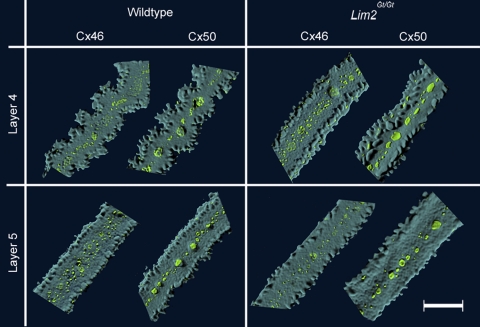
Figure 1.
Fiber cell morphology and gap junctional organization in lenses from wild-type and Lim2Gt/Gt mice. Individual fiber cells were dissected from layers 4 and 5 of the lens and imaged by confocal microscopy. In layer 5 (∼50 μm below the surface), fiber cells from both genotypes were rectilinear in shape and gap junction plaques (green) were restricted to the center of the broad faces of the fiber cells. Further into the lens (layer 4; ∼200 μm below the surface), wild-type fiber cells had an undulating form, whereas Lim2Gt/Gt fibers retained the rectilinear morphology characteristic of the outer (layer 5) cells. Scale bar, 10 μm.

