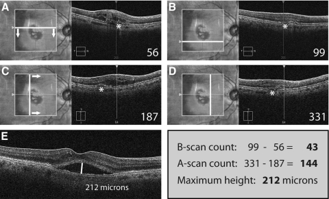
Figure 2.
Simplified grading of SRF in a sample case from the Cirrus HD-OCT. The first B-scan of the cube scan that shows the feature to be graded (SRF marked with an asterisk) is identified and the respective B-scan number noted (A). Then, progressing through the lesion (arrows), the number of the B-scan representing the lower boundary of the lesion feature is also noted (B). In the same way, the A-scans representing the left and right boundaries of the lesion feature are identified and their respective numbers noted. The Cirrus HD-OCT natively supports the A-scan view of a cube scan. Regarding the 3D-OCT-1000 used in our study, we engineered custom software to facilitate an A-scan view similar to that shown for the Cirrus HD-OCT. Finally, the maximum height of the feature is measured with a caliper tool found in virtually any OCT software. The final values for the simplified grading of SRF in this case are: number of B-scans: 99 − 56 = 43; number of A-scans = 331 − 187 = 144; maximum height = 212 μm. Grading of these three parameters will take an average time of approximately 30 seconds per feature.

