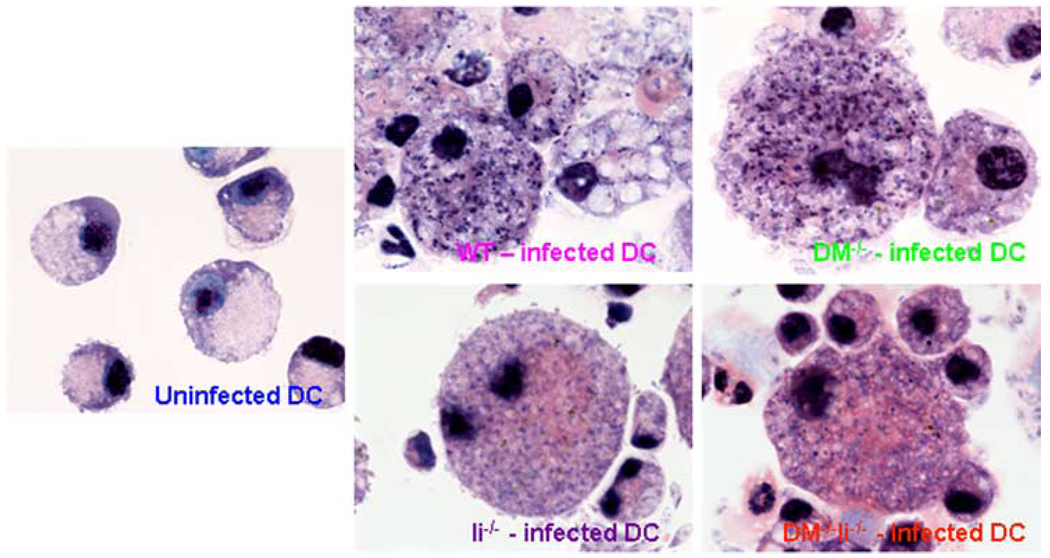
FIGURE 3.
BALB/c wild type, DM−/−, Ii−/−, and DM−/−Ii−/− DC are infected by L. major in a comparable manner. BMDC, prepared as described before (28) and in Materials and Methods, were infected with 35 × 106 L. major promastigotes (at a DC/parasite ratio of 1:35) for 24 h. Cytospin slides prepared from infected and uninfected BMDC were stained with Giemsa (as described in Materials and Methods) and photographed (shown at ×100 magnification). Note that the small black (darkly stained) intracellular dots represent L. major nuclei (and the large black organelle is the DC nucleus).

