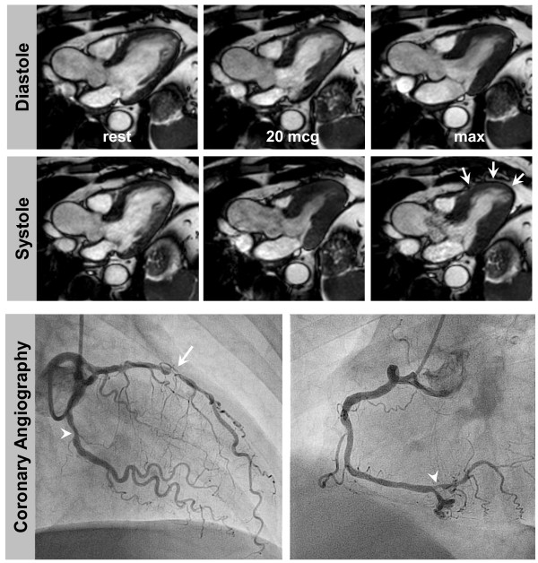
Figure 2.
DCMR in a 56 year old man with exertional dyspnoea and atypical chest pain. He had arterial hypertension and was an active smoker without a prior history of CAD. He was referred for DCMR after a normal exercise ECG and insufficient image quality for a stress echocardiography. DCMR (top and middle) revealed a stress inducible wall motion abnormality of the apical and mid-ventricular anteroseptal segments (white arrows). Invasive angiography (bottom row) demonstrated high grade stenosis of the LAD (white arrow) and intermediate stenoses of the LCX and distal RCA (white arrowheads).

