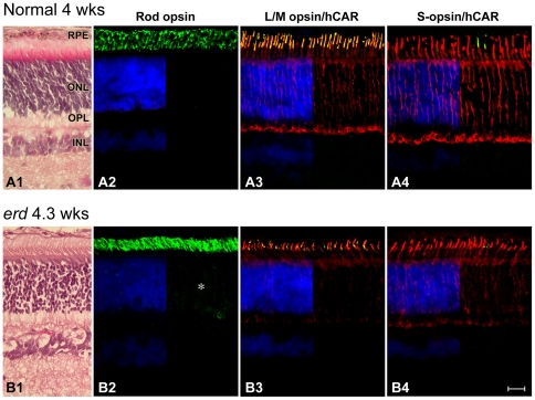
Figure 1. Normal expression of rod and cone molecular markers in early development in erd.
Sections from 4 wk normal (A) and 4.3 wk mutant (B) stained with H&E (A1, B1), or labeled with antibodies against rod opsin (A2, B2; green), L/M-cone opsin (COS-1, green)/hCAR (red) (A3, B3), and S-cone opsin (OS-2, green)/hCAR (red) (A4, B4) with a DAPI (blue) nuclear stain. With the exception of rod opsin which shows slight delocalization of label into the outer nuclear (*) and plexiform layers, opsin labeling is restricted to the outer segments. hCAR labeling of cones is present throughout the cell. RPE = retinal pigment epithelium, ONL = outer nuclear layer, OPL = outer plexiform layer, INL = inner nuclear layer. Scale bar 40 µm.

