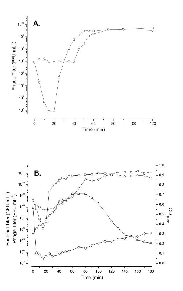
Figure 2.
Effect of multiplicity of infection of phage CBA120 on host cells. 2A. Low MOI (~0.1) single-step growth curve of CBA120 infecting E. coli O157:H7 NCTC 12900. The infection graph shown is representative of three replicates. Infective centers (PFU mL-1, O), and total viable phage (PFU mL-1, □, as determined after the addition of chloroform). 2B. High MOI (~10) CBA120 infection of E. coli O157:H7 NCTC 12900. The infection graph shown is representative of three replicates. Infective centers (PFU mL-1, O), phage (PFU mL-1, □ after the addition of chloroform), optical density (600 nm, Δ) and bacterial titer/survivors (CFU mL-1, ◇).

