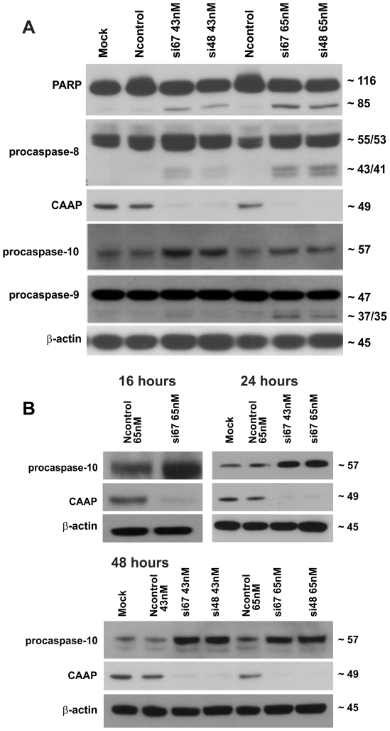
Figure 4. Knockdown of CAAP induces caspase processing, cleavage of PARP and results in increased expression of caspase-10.
(A) Determination of caspase processing and PARP cleavage by Western blot analyses of floating and adherent A-549 cells 48 hours post transfection with si67 and si48 at dosages of 65 nM and 43 nM. The p85 cleavage fragment of PARP after treatment with the siRNAs and the activation of procaspase 9, as shown by the 37/35 fragments, and caspase-8 as shown by the 43/41 fragments, are consistent with the fold increase of Caspase-3, -8, and -9 activities shown in Fig. 3C and discussed in the text. Comparing the expression levels of procaspase 10 in the mock and Ncontrols to the levels in the si67 and si48 lanes implies that the expression of procaspase 10 is increased after RNAi knockdown of CAAP. A representative experiment out of three is shown. (B) Western blot analyses for the status of CAAP and procaspase-10 in floating and adherent A-549 cells 16 h, 24 h or 48 h post transfection with either Ncontrol or si48 or si67 siRNA. Probing the membrane with a ß-actin antibody served as a loading control. Procaspase-10 protein levels were reproducibly higher following knockdown of CAAP.

