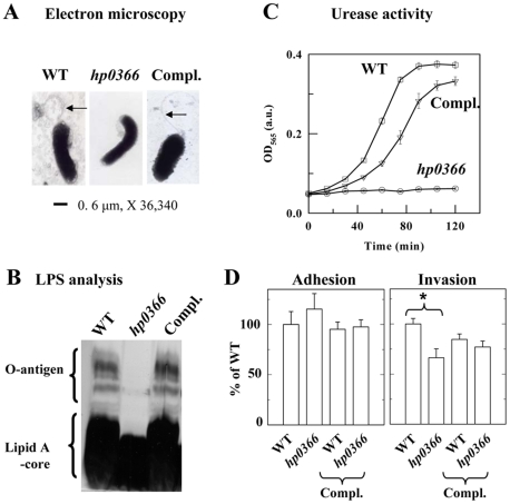
Figure 2. Impact of disruption of hp0366 on virulence factor production and interactions with gastric cells. Panel A:
Analysis of flagellum production by electron microscopy. Arrows point at flagella. Panel B: Analysis of LPS production. LPS was extracted from the WT, mutant and complemented strain (Compl.) and analyzed by SDS-PAGE and silver staining. Panel C: Analysis of the urease activity of WT, mutant and complemented (Compl.) H. pylori. Urease activity was measured using phenol red as an indicator [92]. Experiments were done with 3 different concentrations of soluble protein extracts and the same trend was observed. The data shown were obtained with the highest amount of protein tested and are the average of three replicas. Panel D: Adherence and invasion of H. pylori WT, mutant and complemented strains to gastric AGS cells. The data are expressed in % of adherence or invasion of WT H. pylori. The data are the average of 3 independent experiments. Compl. indicates that the wild-type hp0366 gene was introduced in the strain of interest on a shuttle plasmid. The adherence of WT amounted to 8–10% of the inoculum. No statistical differences (t-test) were observed between WT and mutant strain for adherence. Invasion was measured after elimination of non-internalized bacteria by gentamycin treatment. Invasion represented ∼1.5% of the inoculum for the WT. Statistical differences were observed between the WT and mutant (shown by asterisk, t-test, p<0.001).

