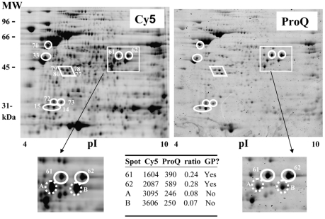
Figure 4. 2D gel electrophoresis analysis of H. pylori glycoproteins using Cy5 labeling of total proteins and ProQ-emerald labeling of glycoproteins.
The proteins from WT H. pylori were stained with Cy5 and were resolved by 2D gel electrophoresis. Glycoproteins were stained with ProQ-emerald. Abundant proteins gave a high background reactivity by ProQ-emerald labeling (ex: UreB). To eliminate false positive proteins, the ratio of the ProQ-emerald and Cy5 signals was calculated and only proteins that showed a high ratio were considered GP candidates. The ratios calculated are indicated for a few spots shown as an example in the zoomed figures. Spots A and B are provided as examples of non glycosylated proteins. Signals for Cy5 and ProQ-emerald are in arbitrary units. Contributions from the gel background have already been subtracted. Note that the analysis was limited to a subset of 100 proteins that were present in sufficient amounts to allow their identification by MS ultimately. The 12 spots highlighted on the figure (in circles and diamond) are the ones with the highest ratios in this subset and represent GPs. Additional GPs may be present.

