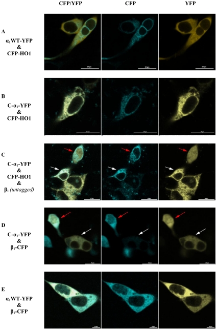
Figure 9. Expression of EYFP-marked α1 full-length (A) and C-α1 (B) in HEK-293 cells.
As a marker of the endoplasmic reticulum [11] ECFP tagged heme oxygenase 1 (human, HO-1) was co-transfected. A, α1 full-length shows cytosolic distribution. B, C-α1 shows a similar distribution like ECFP-HO-1. C, addition of an untagged β1-subunit led in some cells a similar subcellular distribution as the wild type. The red arrow denotes a cell likely co-transfected with β1, the white arrow denotes a cell likely not co-transfected with β1. D, only cells which express both subunits show homogenous subcellular distribution. The red arrow denotes a cell co-transfected with β1, the white arrow denotes a cell not co-transfected with β1. E, The subcellular localization of α1 full-length is not affected through coexpression of β1. Bar, 20 µm. CFP, ECFP channel; YFP, EYFP channel.

