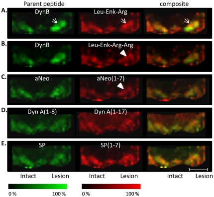
Figure 7. MALDI IMS ion images of peptides.
(A) Composite MALDI IMS ion images from one single section of a high-dyskinetic animal reveal regional a distinct regional discrepancy between locally low Dyn B (green) and corresponding high Leu-Enk-Arg (red) levels in the dorsolateral SN (arrow). (B, C) Dynorphin metabolites Leu-Enk-Arg-Arg and aNeo(1–7) were also upregulated in the lateral, but not medial part of SN in HD animals (arrowhead). (D) Nigral levels of DynA (1–8), DynA (1–17), and Substance P (E) were not associated with any treatment-induced changes. Scale bar = 2 mm.

