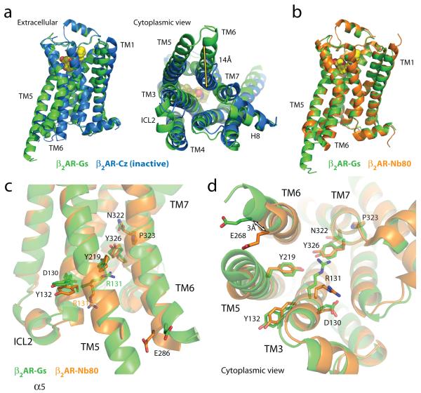
Figure 3. Comparison of active and inactive β2AR structures.
a, Side and cytoplasmic views of the β2AR-Gs structure (green) compared to the inactive carazolol-bound β2 AR structure 3 (blue). Significant structural changes are seen for the intracellular domains of TM5 and TM6. TM5 is extended by two helical turns while TM6 is moved outward by 14 Å as measured at the α-carbons of Glu268 (yellow arrow) in the two structures. b, β2AR-Gs compared with the nanobody-stabilized active state β2AR-Nb80 structure 12 (orange). c, The positions of residues in the E/DRY and NPxxY motifs and other key residues of the β2AR-Gs and β2AR-Nb80 structures. All residues occupy very similar positions except Arg131 which in the β2AR-Nb80 structure interacts with the nanobody. d, View from the cytoplasmic side of residues shown in (c).

