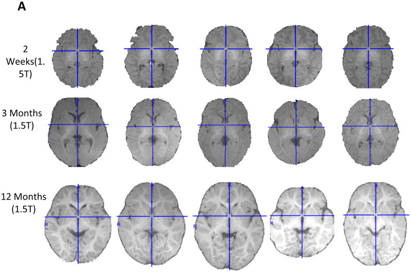
Figure 2.
Axial slice of extracted brain for individual infants at 2 weeks, 3, 12, or 18 months, or 4 years of age for 1.5T (2A), or 3 and 6 months of age for 3T (2B). The crosshairs are placed on the anterior commissure (AC) and run through the posterior commissure (PC). The individual figures preserve the relative size of the brain for children within one age-group, and the differences across age retain the approximate relative size of the head. Note: all axial slices shown in figures are oriented to the Talairach stereotaxic space, which places a line drawn from the AC to the PC orthogonal to the sides of the MRI volume, and is the approximate alignment of the MNI-152 average template (. All axial MRI volumes in all figures are shown with the left side of the brain on the right side of the figure.


