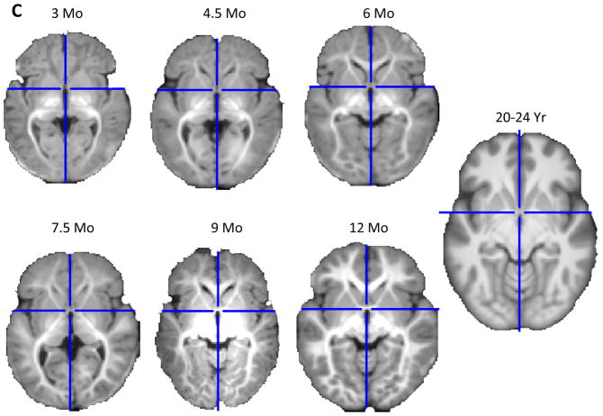
Figure 3.
Age-specific templates for 1.5T scans showing mid-sagittal slice (A) and axial slice at AC-PC commissure (B) of whole head average template T1W MRI volumes across ages of study. The individual figures preserve the relative size of the head for children at that age. Age-specific templates for 3T scans showing axial slice (C) of brain average template T1W MRI volumes. Note: all axial slices shown in figures are at the origin location of the Talairach stereotaxic space, which is a line drawn from the anterior commissure to the posterior commissure. The scans come from 1.5T scans for 2 weeks, 3, 6, 9, 12 months, 1.5, 2, 2.5, 3, and 4 yrs; 3T scans for 3, 4.5, 6, 7.5, 9, and 12 months. The 20–24 year scans come from participants aged 20 through 24.9 years and are combined 1.5T and 3T scans (3A, 3B) or only 3T scans (3C).


