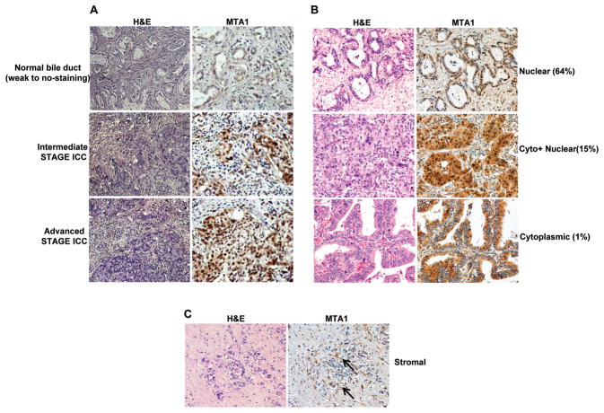
Figure 6.
Evaluation of MTA1 expression in O. viverrini-induced human cholangiocarcinomas. (A) Immunohistochemical analysis of MTA1 in bile duct hyperplasia, early stage cholangiocarcinoma, and advanced-stage cholangiocarcinoma. (B) Representative examples of subcellular localization of MTA1 in the nuclear, cytolasmic, and both compartments (Cyto + Nuclear). (C) Representative examples of MTA1 immunoreactivity in the stromal compartment; arrowed indicate examples of MTA1-positive, stromal fibroblasts. For each situation, consecutive paraffin sections were stained with Haematoxylin and Eosin (H&E) (left panels) and with anti MTA1 antibody (right panels).

