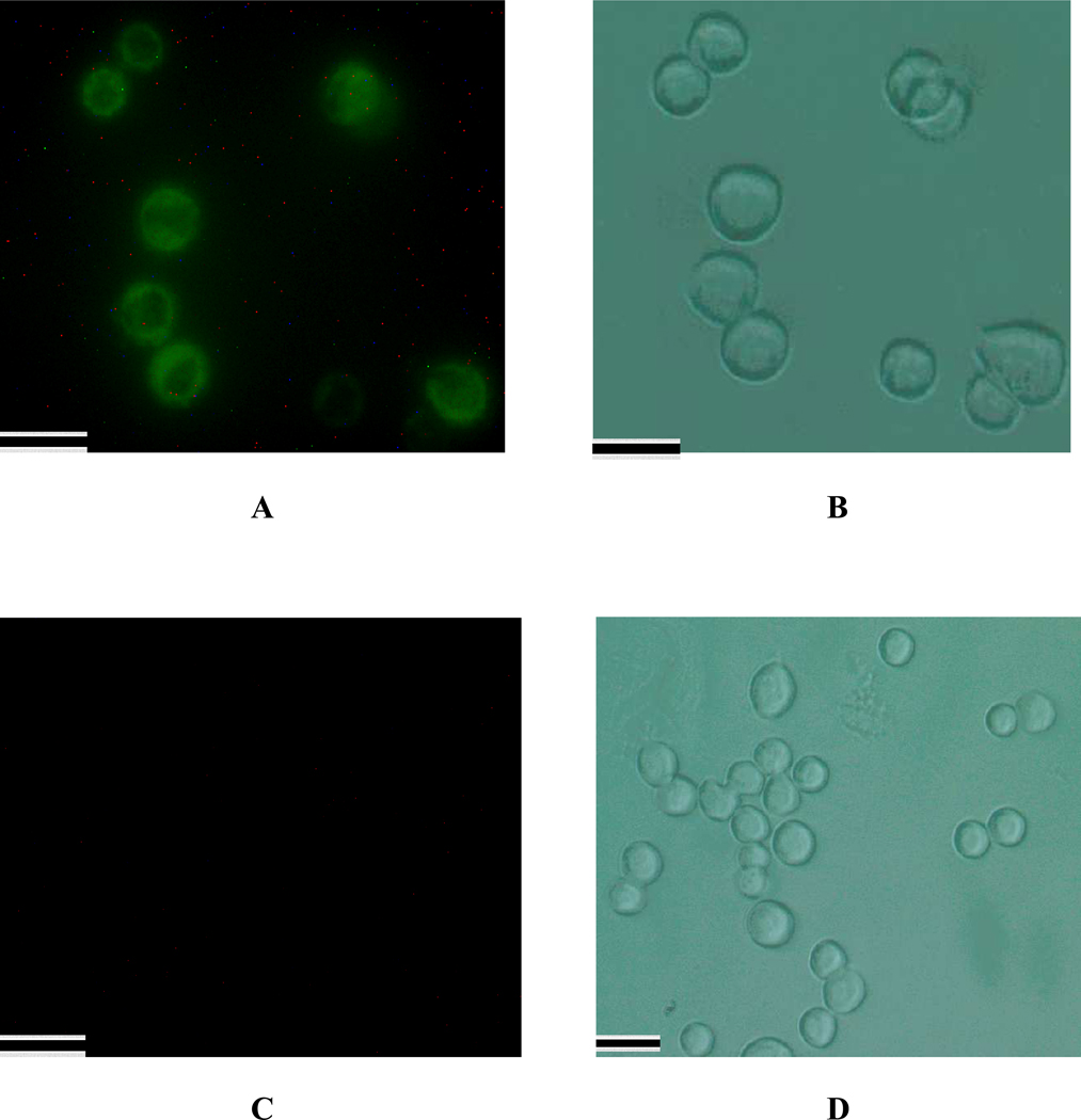
Figure 5.
SK-BR-3 cancer cell fluorescent images after incubation with Cy3 modified S6 aptamer attached GNPOP modified SWCNT for an hour. Green indicates the presence of Cy3 labeled nanomaterial on the cell. B) Bright-field image of SK-BR-3 cells after incubation with Cy3 modified S6 aptamer attached GNPOP modified SWCNT. (C) MDA-AB cell fluorescent images after incubation with Cy3 modified S6 aptamer attached GNPOP modified SWCNT for 4 hours. No green fluorescence was observed. D) Bright-field image of MDA-AB cells after incubation with Cy3 modified S6 aptamer attached GNPOP modified SWCNT. Scale bar represents 20 µm.

