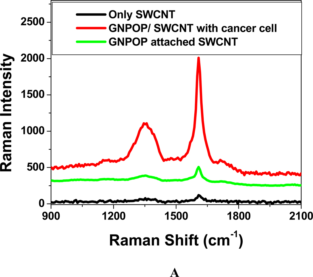
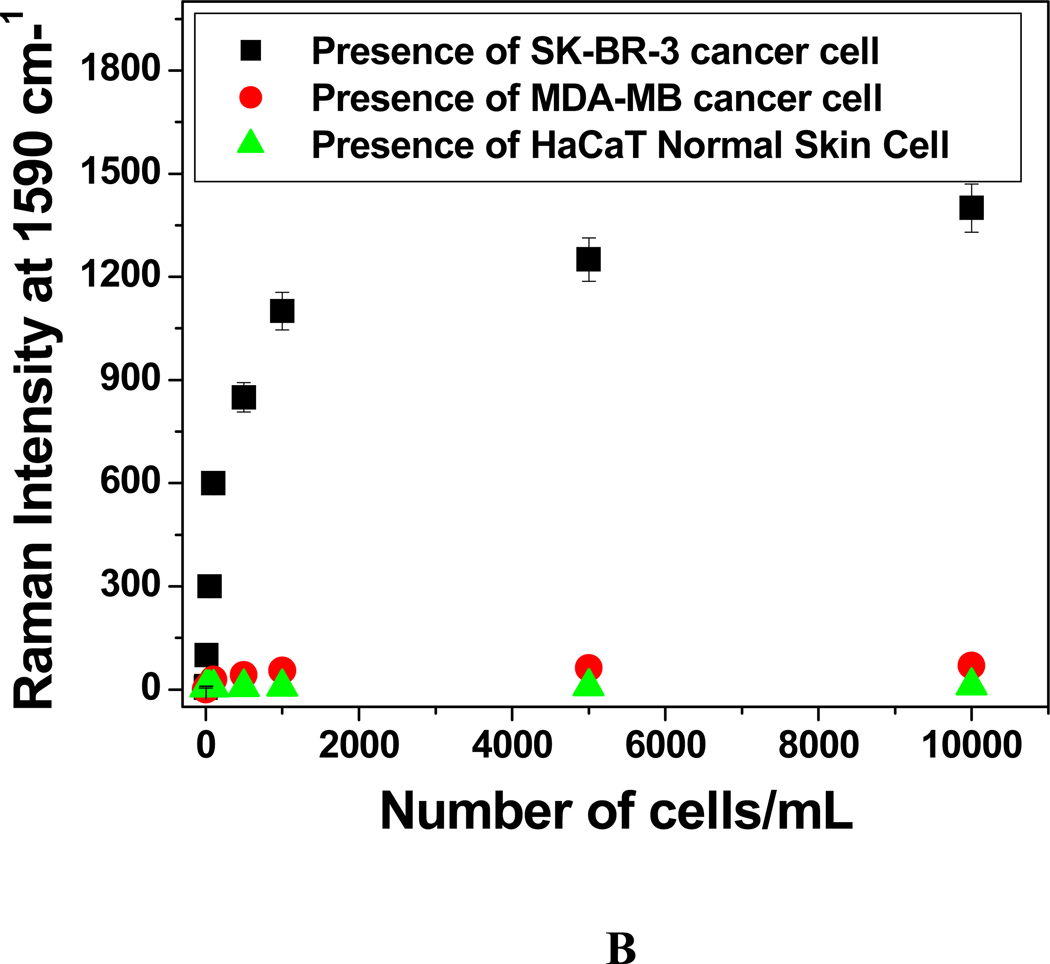
Figure 6.
A: Plot showing SERS enhancement in the presence of 104 cells/mL SK-BR-3 breast cancer cell. B) Plot showing SERS scattering intensity change at 1590 cm−1 upon the addition of different concentrations (number of cells/mL) of SK-BR-3, MDA-MB human breast cancerous and HaCaT normal skin cells to S6 aptamer conjugated GNPOP attached SWCNTs. In all cases, we have used 10 µg/mL SWCNT and 10 µg/mL SWCNT hybrid.


