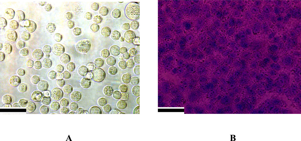
Figure 7.
A) Bright field inverted microscope images of aptamer conjugated hybrid nanomaterial attached SK-BR-3 breast cancer cells stained with trypan blue, before irradiation. B) Bright field inverted microscope images after irradiation with 785 nm light with power 1.5 W/cm2 for 10 minutes followed by staining with trypan blue. Scale bar represents 20 µm.

