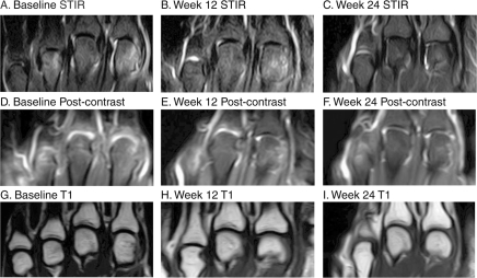
Figure 3.
MRI of the wrist at baseline (A, D, G), week 12 (B, E, H) and week 24 (C, F, I) of a patient randomised to receive golimumab 100 mg plus placebo. Coronal short tau inversion recovery (STIR) images (A–C) show extensive bone oedema at baseline (A). The bone oedema has markedly decreased at week 12 (B) and has nearly resolved at week 24 (C). Corresponding postcontrast T1-weighted images with fat suppression (D–F) show substantial synovitis at baseline (D) and markedly reduced synovitis at week 12 (E) and week 24 (F). Precontrast T1-weighted images without fat suppression (G–I) show no progression of bone erosion during the 24-week follow-up period. Note: Series of consecutive images were evaluated; the images displayed here are representative but not exhaustive.

