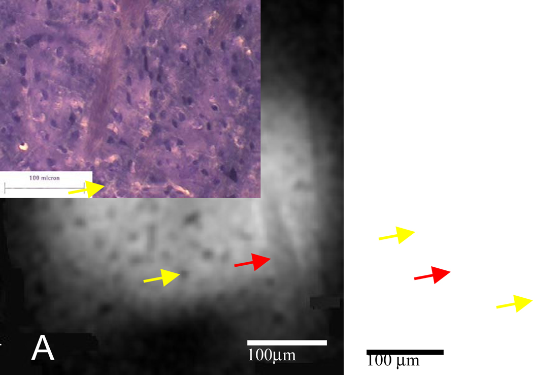
Figure 2.
MR microscopy and histology of rat striatal tissue. (A) Representative MR image (4.7µm isotropic) from a three-dimensional gradient echo data set acquired at 4.7µm isotropic resolution (TE=10ms, TR=150ms, matrix=1283, 0.6mm FOV, acquisition time=22h) with (B) histology from a similar piece of tissue. The image and histology show descending white matter tracts (red arrows). Structures which resemble cell bodies of medium spiny neurons in size, shape, and distribution are present throughout (yellow arrows show two examples).

