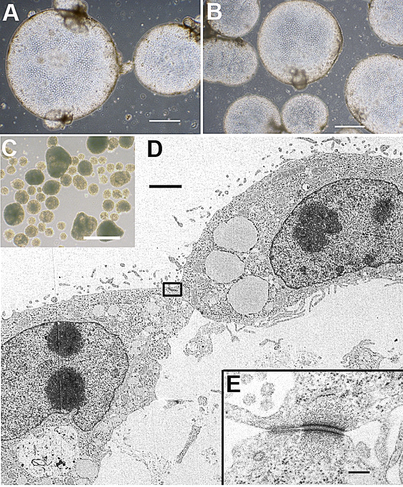
FIG. 5.

Cell sphere formation from iTR cells is shown. Spheres from iTR1 p42 (A) and iTR2 p7 cells are shown (B). C) Embryoid body formation from an iPS cell line (ID6, p5) is shown. Bars in A–C = 500 μm. D) Ultrastructure of a portion of a cell sphere from iTR2 cells (85-nm-thick section; bar = 2 μm). E) Cell junction (area boxed in D) shows a desmosome-like structure. Bar = 0.2 μm.
