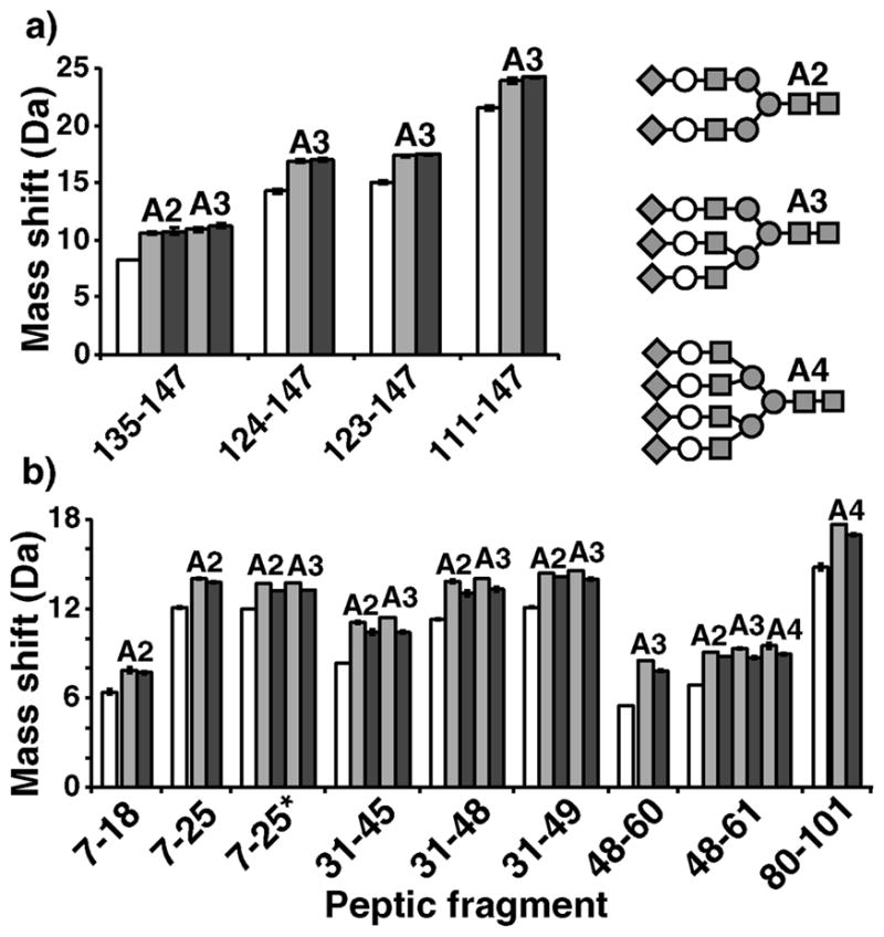
Figure 4.

Glycopeptide deuteration data on Fetuin (a) and human α1AGP (b). Mass shifts from after exhaustive deuteration are shown for various deglycosylated (PNGaseF treated) (white), asialo (Neuase treated) (light grey), and fully sialylated (untreated) (dark grey) peptic fragments of each protein. Labels above bars indicate degree of branching: biantennary (A2), triantennary (A3), and tetraantennary (A4). *Fragment 7–25 of α1AGP displayed two variants containing either Arg or Gln at position 20.
