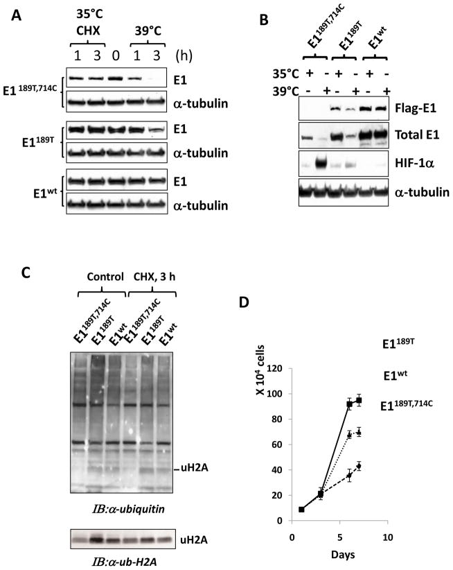
Fig. 5. Stable expression of 714C to W revertant restores E1 function.
A: Two newly established cell lines E1189T and E1wt together with their TS20 cell line, which contains E1189T,714C mutations, were analyzed. Cells were culture at permissive 35°C or restrictive 39°C for 1 or 3 h. Cycloheximide (Sigma) were used to block de novo translation and test the protein stability. Total E1 protein levels were detected by using an anti-UBE1 antibody. α-tubulin was detected as a loading control by using an anti-α-tubulin antibody. Note that in stable transfected cell lines, the exogenous E1 represent the major part of total E1. B: The three cell lines used in A were cultured at both temperature and restrictive temperature points. Flag-E1 and total E1 protein levels were detected using anti-flag and anti-UBE1 antibodies, respectively. Ubiquitin proteasome pathway targeted protein, HIF-1α level was determined as an indicator of the polyubiquitination-proteasomal function while α-tubulin levels were examined as a loading control. C: Ubiquitination profile in the three cell lines were examined by using anti-ubiquitin antibody, and a band representing monoubiquitinated H2A (uH2A) was indicated. Its identity was further confirmed by anti-Ub-H2A specific antibody. D: The proliferation rates of the three cell lines were assayed at permissive temperature. Cell numbers (determined by DNA contents) were determined at days 0, 1, 3, 6 and 7. Standard errors were indicated.

