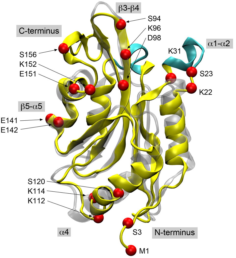
Figure 2.
A structural alignment of human cofilin (yellow) and yeast cofilin (silver). The secondary structure elements discussed in the text are the shaded labels and the locations of lethal point mutations explained by our docking model are shown as red spheres and labeled based on the human cofilin sequence. The major loop insertions in the human protein are highlighted in cyan.

