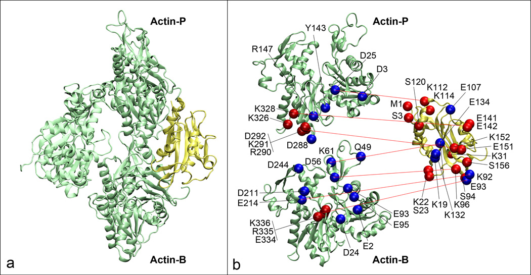
Figure 3.
Molecular details of the interaction between cofilin and the actin filament. (a) An actin trimer (green) where we have labeled the two actin protomers, Actin-P and Actin-B that contact cofilin (yellow). (b) Molecular details where lethal mutations for both proteins are shown as red spheres while other contact points predicted by the model are shown in blue and red lines indicate contact points. The two actin protomers have been spread apart to more clearly show the interactions.

