Figure 1.
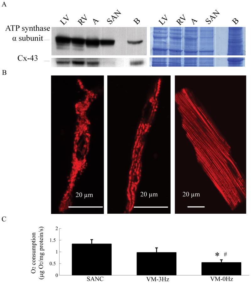
Sinoatrial node cells (SANC) have a high density of mitochondrial proteins and a high basal respiratory rate: (A) Immunoblotting of the mitochondrial complex V and connexin 43 in rabbit tissues; LV-left ventricle, RV-right ventricle, SAN-sinoatrial node and B-brain. RV, LV and SAN have mitochondrial protein densities similar to other tissues. (Lack of expression of gap junction protein, connexin 43 verifies that the SAN tissue is truly SAN, and not atrial tissue). Colloidal blue staining of the gel in the right panel confirmed comparable protein amounts among all the samples loaded on the gel. (B) SANC (left and middle) and VM (right) mitochondria visualized by tetramethylrhodamine methyl ester staining. Colloidal blue staining of the gel in the right panel confirmed comparable protein amounts among all the samples loaded on the gel. (C) Respiration rates of isolated spontaneously beating SANC suspension (n=17) and quiescent or electrically stimulated (3Hz) ventricular myocytes (VM) suspension (n=8). *p<0.05 vs. SANC, #p<0.05 vs. VM-3Hz.

