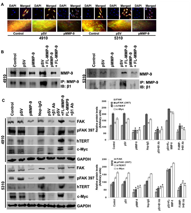
Figure 5. MMP-9 silencing inhibits β1-integrin-mediated hTERT expression in glioma cells.
A. pMMP-9-transfected glioma cells were analyzed by immunocytochemistry for co-localization (yellow color) of MMP-9 (green color) and β1-integrin (red color) using specific antibodies. B. After 24 hours of pMMP-9 transfection, 4910 and 5310 glioma cells were double transfected with FL-MMP-9 and incubated for 24 hours. Western blot analysis shows effect of FL-MMP-9 transfection on MMP-9 expression in 4910 and 5310 glioma xenograft cells compared to control and pSV. Immnunoprecipitation was carried out by anti-MMP-9 antibody and immuprobed with β1-integrin. C. After 24 hours of pMMP-9 transfection, 4910 and 5310 glioma cells were double transfected with FL-MMP-9, non-specific IgG and/or anti-β1-integrin antibodies for another 24 hours. Western blot analysis was performed for FAK, phospho FAK (Tyr 397), hTERT and c-Myc using total cell lysates. GAPDH served as loading control. Protein band intensities were quantified by densitometric analysis using ImageJ software (National Institutes of Health). Each bar represents triplicate analyses of mean ± SD. Significant changes are represented by an asterisk (*) (P <0.05). (n=3).

