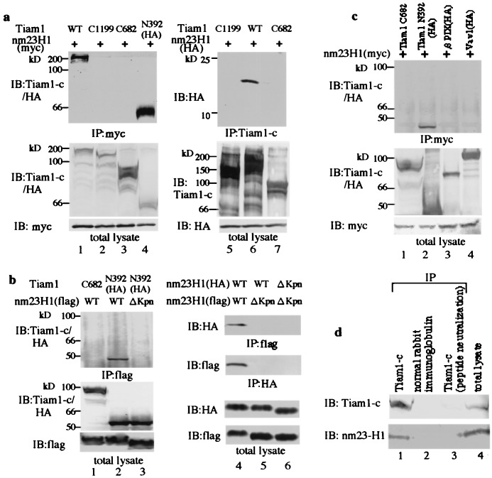
Figure 2.
Nm23H1 specifically associates with the guanine nucleotide exchanging factor Tiam1. (a) 293T cells were transiently transfected with plasmid encoding myc or HA-tagged nm23H1 together with the Tiam1 constructs indicated above the lanes. Cells were lysed and immunoprecipitated (IP) with anti-myc (lanes 1–4) or anti-Tiam1-c (lanes 5–7), which reacts with epitopes located at the carboxyl terminus of Tiam1. Bound proteins were immunoblotted (IB) with the antibodies indicated. Expression of Tiam1 and nm23H1 in individual cell lysates (total lysate) was confirmed by immunoblotting (Bottom). (b) 293T cells were transiently cotransfected with flag-tagged wild type or ΔKpn nm23H1 together with Tiam1 constructs as indicated. Cell lysates were immunoprecipitated with anti-flag and immunoblotted with the antibodies indicated (lanes 1–3). HA-tagged wild-type or ΔKpn nm23H1 constructs were transiently transfected with flag-tagged wild-type or ΔKpn nm23H1 constructs (lanes 4–6). Immunoprecipitation and immunoblotting were performed as indicated. Expression of nm23H1 and Tiam1 is shown at the bottom. (c) 293T cells were transiently transfected with plasmid encoding myc-tagged nm23H1 together with Tiam1, HA-tagged βPIX, or Vav1 as indicated. Cell lysates were immunoprecipitated with anti-myc and immunoblotted sequentially with anti-Tiam1-c and anti-HA antibodies. (d) Whole brains of adult Institute for Cancer Research (ICR) mice were lysed and immunoprecipitated with anti-Tiam1-c antibody, normal rabbit Ig fraction, or peptide-neutralized anti-Tiam1-c antibody. Precipitants were subjected to immunoblot with the indicated antibodies. These experiments were performed at least three times.

