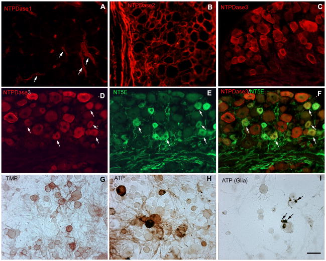
Fig. 4. Restricted distribution of NTPDase immunoreactivity in DRG.
(A–B) Immunohistochemical staining of lumbar DRG for NTPDases shows only vascular staining for NTPDase1 (arrows in A), while NTPDase2 staining is limited to glia. (C) NTPDase3 labeled a substantial subset of neurons including both small (likely unmyelinated) and large (likely myelinated) neurons. (D–F) Double labeling of lumbar DRG indicates most NT5E-positive cells (green) are also labeled for NTPDase3 (red). Arrows show positive cells in (D) and (E), arrows show double labeled cells in (F). (G– H) Histochemical staining of dissociated mouse DRG neurons using TMP (3 mM) and ATP (1 mM) as substrates to confirm neuronal labeling. Neurons labeled for AMPase activity were principally small, whereas ATPase activity labeled both small and large cells. (I) Glial cells were also labeled in the presence of ATP (arrows). Scale bars: A–I, 50 uM.

