
Figure 3.
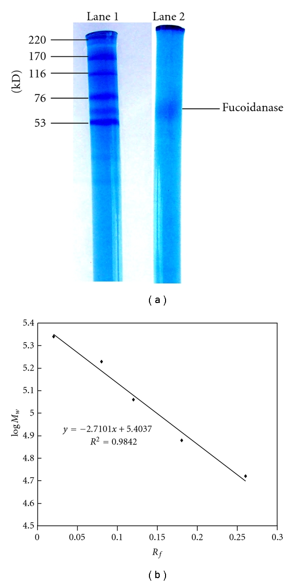
Determination of the M w of the fucoidanase by SDS-PAGE. Picture of electrophoresis of fucoidanase: lane 1 is M w marker; lane 2 is the fucoidanase; (b) standard curve and M w function of the M w marker SDS-PAGE was conducted using discontinuous electrophoresis method with Phastsystem, 5.5% (w/v) polyacrylamide, pH 8.3, 250 V, 10 mA, in the concentration gel with 7.0% polyacrylamide (w/v), 250 V, 30 mA, in the separation gel. Protein was stained with Coomassie brilliant blue R-250. Molecular markers used were (220 kDa), α-2 macroglobulin (170 kDa), β-galactosidase (116 kDa), transferring (76 kDa), and glutamic dehydrogenase (53 kDa).