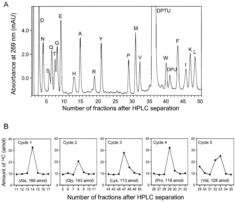
Figure 3.
(A) Typical UV trace of random peptide-bead internal standard (7 beads) and carrier protein, β-lactoglobulin (50 pmol) co-chromatographing with AMS detection. (B) Amount of 14C measured by AMS in HPLC fractions collected every 30 sec manually of 453 amol [14C]GST sequence analysis. The values in parentheses are yields calculated as described in the text.

