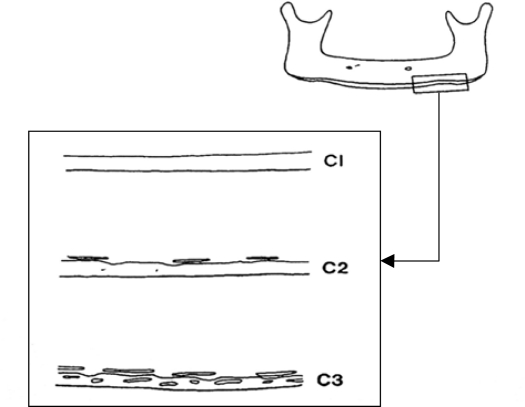
Fig 1.
Diagram illustrating the classification of the endosteal inferior cortex on panoramic radiographs.
C1: The endosteal margin of the cortex is even and sharp on both sides of the mandible.
C2: The endosteal margin has semilunar defects (resorption cavities) with cortical residues one to three layers thick on one or both sides.
C3: The endosteal margin consists of thick cortical residues and is clearly porous.

