Abstract
Introduction:
Esthetics and attractiveness of the smile is one of the major demands in contemporary orthodontic treatment. In order to improve a smile design, it is necessary to record “posed smile” as an intentional, non-pressure, static, natural and reproducible smile. The record then should be analyzed to determine its characteristics. In this study, we intended to design and introduce a software to analyze the smile rapidly and precisely in order to produce an attractive smile for the patients.
Materials and Methods:
For this purpose, a practical study was performed to design multimedia software “Smile Analysis” which can receive patients’ photographs and videographs. After giving records to the software, the operator should mark the points and lines which are displayed on the system’s guide and also define the correct scale for each image. Thirty-three variables are measured by the software and displayed on the report page. Reliability of measurements in both image and video was significantly high (α=0.7–1).
Results:
In order to evaluate intra- operator and inter-operator reliability, five cases were selected randomly. Statistical analysis showed that calculations performed in smile analysis software were both valid and highly reliable (for both video and photo).
Conclusion:
The results obtained from smile analysis could be used in diagnosis, treatment planning and evaluation of the treatment progress.
Keywords: Analysis, Esthetics, Orthodontics, Software
INTRODUCTION
Since the introduction of orthodontics as a separate specialty in dentistry, a variety of events have happened in its methods, instruments and treatment plans. In the 20th century Angle’s paradigm with the priority of ideal occlusion and correction of the skeletal orthodontic treatment.
According this relation of the jaws had become the essence of paradigm, if occlusion is corrected as he defined, then the soft tissue will be fine. But these changes do not always happen and if they happen they will not always be in line of improvement [1–5]. At the beginning of the 21th century, an intention to soft tissue paradigm became the base of diagnosis and treatment planning in orthodontics [3–6]. According to this paradigm, nothing was more important and functional than facial esthetics in dentistry [5] and obtaining ideal esthetics and harmony in the oral and facial soft tissue was the first aim in orthodontic treatment [1,4]. Apparent esthetics, especially facial esthetics, has an important role in confidence and acceptance of the patient in public [4,7–9]. The way people contact each other is under the phenomenon “attractiveness halo” in which people have more positive acceptance and better behavior against attractive faces [8]. These factors made obtaining an ideal esthetic the most common demand for orthodontic treatment. Among parts of the face, eyes have the most important role in esthetics and the mouth (especially when smiling) is afterwards [8–10]. Having a beautiful smile may decrease the negative effect of “attractiveness halo” in non-so-attractive people [8].
There are different classifications for the smile in clinical implication which are different fundamentally, but the most practical classification for the evaluation of a smile was presented by Ackerman and Ackerman which was accordingly divided into two groups of “posed smile” and “unposed smile” [4,11,12].
Posed smile is an intentional informed smile which is without any stimulations or emotions; it is static, well presentable and prolonged like when a person wants to take a photograph. This smile can be strained or non-strained.
On the other hand, “unposed smile” (enjoyment smile) is a non intentional and spontaneous smile which is induced by emotional factors like happiness and excitement [4,11,12]. Different studies show that an unstrained posed smile is the best smile for recording and analyzing in orthodontics and other fields of dentistry [4,12–15]. Records required for integrative smile analysis include frontal at rest, frontal smile, profile, oblique smile and frontal and oblique close-up smile views which can be obtained statically (photography) or dynamically (videography). One of the advantages of videography is providing a wider range of images which enables the clinician to choose the best image of the posed smile [16]. Moreover, the state of the patient’s speech and smile and his oral and pharyngeal function can be recorded at the same time [6,17]. After providing static and dynamic records, the clinician could perform the required analysis over the resulted data to use them practically in his diagnosis and treatment plan. For this, at first he should find factors needed and then using available instruments measure them from his patients’ records. For full smile analysis it is necessary to evaluate the factors in four dimensions; namely, frontal, sagittal, oblique and the fourth dimension which is time. In each dimension some of the related variables could be measured. In the frontal view, the lip length and lip curvature, vertical relationship of the teeth and lips, transverse dimension of the dental arch and its relation with the facial width, vertical and horizontal symmetry of the smile, transverse cant of the occlusal plan and other dimensions are measured. In the sagittal view, incisor inclination and overjet are measured and the most important factors in the oblique view are the smile arc and anteroposterior cant of the occlusal plan. Time dimension explains changes which have occurred in smile characteristics during growth and adolescence.
Then, these factors should be evaluated in the records of patients. Hulsey appointed a series of reference lines on smile photographs of his 40 cases and calculated five parameters in each photo [13]. Other studies used direct measurements on the photos and later digital calculations. Ackerman et al in 1998 designed “smile mesh” which was a software for performing analysis. In its primary version, the operator should place a framework consisted of three horizontal and four vertical lines over the close-up photography, then the software measured 11 proportions in the smile [12]. In the next versions, the number of lines increased to five horizontal and seven vertical lines and the reported variables increased to 16 [4]. After presenting this software, some studies used it in their analysis [6,17]. Limitations in its use like the high price and also limitation in using dynamic records made us provide the software with more facilities to benefit it in diagnosis, treatment planning and evaluation of treatment results and also in research fields around smile.
MATERIALS AND METHODS
The first step in designing any software is gathering information and finding the required variables. Reviewing available texts and references, variables associated with smile were collected, the most precise and practical definition for each variable was chosen and the necessary modification was carried out in some cases. These variables and their scientific-practical definition are listed in Table I. Then, the software “smile analysis” was prepared with coding language “Visual Basic 6” under information bank ACCESS.
Table I.
List of variables used in smile analysis and their definitions
| Variable | Practical definition |
|---|---|
| Philtrum height at rest | The distance between subspinale and inferior point of upper lip vermilion under philtrum column at rest |
| Commissure height at rest | The distance between the line passed from subspinale parallel to alar base to outer commissures |
| Upper lip curvature at rest | Upper lip curve from midline to outer commissures at rest |
| Interlabial gap at rest | The distance between inferior upper lip border and superior lower lip border at rest |
| Upper Incisor exposure at rest | The amount of upper incisors exposure at rest |
| Lower incisor exposure at rest | The amount of lower incisors exposure at rest |
| Philtrum height on smile | the distance between subspinale and inferior point of upper lip vermilion under philtrum column on smile |
| Commissure height on smile | The distance between the line passed from subspinale parallel to alar base to outer commissures on smile |
| Upper lip curvature on smile | Upper lip curve from midline to outer commissures on smile |
| Interlabial gap on smile | The distance between inferior upper lip border and superior lower lip border on smile |
| Upper incisor exposure on smile | The amount of upper incisors exposure on smile |
| Lower incisor exposure on smile | The amount of lower incisors exposure on smile |
| Smile index | The result of smile width divided by interlabial gap |
| Intercommissure width (smile width) | The distance between outer commissures on smile |
| Buccal corridor | The distant between the most distal point of upper right canine or first premolar to inner commissure on smile |
| Buccal corridor ratio | The result of distance between lines tangent on most distal points of upper canines or first premolars divided by smile width on smile |
| Gingival display | Amount of gingival display of upper central incisors from marginal gingiva to inferior border of upper lip on smile |
| Transverse symmetry | Smile symmetry in transverse dimension according to differential distance between outer commissures and midline in both sides |
| Vertical symmetry | Smile symmetry in vertical dimension according to the angle between line passes through outer commissures and midline |
| Transverse cant of occlusal plane | Cant of occlusal plane (line passes through peaks of upper canines) according to its angle with midline |
| Upper dental midline angulation | Angle between upper dental midline and facial midline |
| Upper dental midline deviation | Distance between upper dental midline and facial midline and its direction |
| Over jet | Distance between labial surface of upper and lower central incisors |
| Incisor inclination | Anterior-posterior inclination of longidudinal axis of upper central incisors in smile sagittal view in relation to frontal plane |
| Smile arc | The difference between distance of incisor edge of upper central incisors, and peak of upper right and left canines or first premolars to inferior border of upper lip |
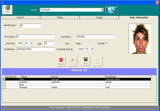
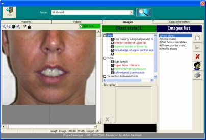
The program includes four sections; namely, “basic information”, “images”, “videos” and “reports”. After entering the information of each patient in the basic information section (Fig 1), the operator can place smile records of each patient (including images or videos) in the associated part. Then, the operator should place points and lines on the available records according to the sample existing in the guide section of the program (Fig 2). Using high magnification at the time of placing points and lines predominantly increases the accuracy.
Fig 1.
The section “basic information
Fig 2.
The section “images”
For reducing problems due to different magnification in images, it is possible to determine the scale of each image independently. In order to do that, the operator should measure the distance between two imaginary points on an image and compare it with the real size in the patient’s face or model; so, all calculations can be performed according to the new-determined scale. For optimal use of this facility, it is recommended to place a scaled device such as a ruler at the same level to the patient’s face around him/her when preparing records in order to increase the precision of calculations. In “images” section, it is possible to determine the distance between two favorite points (other than that referred in reports). This facility is especially helpful in performing other soft tissue analyses. It is also possible to superimpose images on the screen: after selecting two images and moving the scroll bar to the right, the first image is gradually obliterated and the second image appears on it. This provides a very helpful tool for superimposing lateral cephalometries and profile photos of the patients. However, it is necessary to determine standard measures in taking extra-oral photographs in order to enable superimposition appropriately. This part is very helpful in evaluating facial movements from the rest position to gestic smile and subsequently the complete (enjoyment) smile especially in the surgical field for rehabilitation of inabilities due to facial nerve palsy.
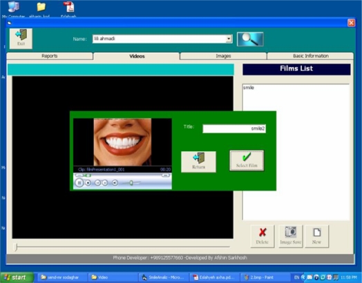
The operator could place the videos in different views (frontal, profile, oblique) in full-face or close-up in “film” section and after necessary evaluation, choose his favorite frame as a separate image, save it in the “images” section, and analyze it as a usual image (Fig 3).
Fig 3.
The section “films”
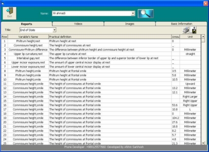
So images that could be analyzed are selected from the patient’s image file or from the patient’s videos after capturing the favorite image. In the “reports” section, 32 smile-associated variables, each measured in one of the five views (rest, frontal smile, close-up smile, profile and oblique smile) are displayed with their definitions (Fig 4).
Fig 4.
The section “reports”
RESULTS
The software can be downloaded free from the following website: http://drc.tums.ac.ir/content/?contentID=115
In order to test the software and evaluate its reliability based on the only available study [12], five cases were selected randomly.
To prepare the images, the samples were placed in the natural head position (NHP).
To prepare these videographs, a digital camer(SONY P73, 4.1 MP) was placed in 80cm distance of sample so that the camera lens was parallel to patient’s face –in NHP- and level to inferior third of the face.
To omit the problems of different magnification, a scaled ruler placed near the patient’s face and level to his mouth was used as a scale.
Then, two trained dental students as operators evaluated the records with “smile analysis” software.
After a week, records were again delivered to the operators, in absolutely similar manner, for reevaluation.
After collecting data, they were statistically analyzed with SPSS 14.
The obtained results of intra- operator and inter-operator reliability are summarized in table II and table III for photographs and table IV for videographs.
Table II.
Reliability of calculations on photographs in quantitative variables
| Variable |
RELIABILITY |
||
|---|---|---|---|
| Operator A | Operator B | Inter-operator | |
| Philtrum height, rest | 0.965 | 0.984 | 0.987 |
| Commissure height, rest | 0.991 | 0.996 | 0.995 |
| Philtrum, commissure difference, rest | 0.996 | 0.996 | 0.998 |
| Interlabial gap, rest | 0.993 | 0.999 | 0.985 |
| Upper incisor exposure, rest | 0.997 | 0.998 | 0.998 |
| Philtrum height, smile | 0.968 | 0.951 | 0.997 |
| Commissure height, smile | 0.997 | 0.937 | 0.885 |
| Philtrum, commissure difference, smile | 0.995 | 0.991 | 0.932 |
| Interlabial gap, smile | 0.983 | 0.981 | 0.967 |
| Upper incisor exposure, smile | 0.975 | 0.974 | 0.993 |
| Lower incisor exposure, smile | 1 | 0.997 | 0.975 |
| Smile symmetry, right | 0.942 | 0.990 | 0.961 |
| Smile symmetry, left | 0.970 | 0.970 | 0.966 |
| Smile width | 0.989 | 0.992 | 0.986 |
| Smile index | 0.995 | 0.997 | 0.970 |
| Buccal corridor, right | 0.962 | 0.973 | 0.963 |
| Buccal corridor, left | 0.782 | 0.980 | 0.929 |
| Buccal corridor ratio | 0.870 | 0.862 | 0.902 |
| Incisor inclination | 0.913 | 0.943 | 0.824 |
| overjet | 0.987 | 0.993 | 0.994 |
| Upper dental midline angulation | 0.960 | 0.998 | 0.953 |
| Upper dental midline deviation | 0.980 | 0.980 | 0.960 |
| Gingival display, smile | 0.997 | 0.996 | 0.994 |
Table III.
Reliability of calculations on photographs in qualitative variables
| Variable |
RELIABILITY (percent) |
||
|---|---|---|---|
| Operator A | Operator B | Inter-operator | |
| Upper lip curvature, rest | 100 | 100 | 100 |
| Upper lip curvature, smile | 100 | 100 | 100 |
| Cant of occlusal plan | 100 | 100 | 100 |
| Transverse symmetry | 75 | 50 | 60 |
| Vertical symmetry | 100 | 100 | 80 |
| Smile arc | 100 | 100 | 100 |
Table IV.
Reliability of calculations on videograph
| Variable | RELIABILITY |
|---|---|
| Interlabial gap, smile | 0.947 |
| Upper incisor exposure, smile | 0.939 |
| Lower incisor exposure, smile | 0.992 |
| Smile width | 0.955 |
| Smile index | 0.902 |
| Buccal corridor, right | 0.696 |
| Buccal corridor, left | 0.809 |
| Buccal corridor ratio | 0.449 |
| Gingival display, smile | 0.838 |
Since the qualitative variables could not be evaluated by SPSS, the percent of intra-operator and inter-operator similarity was reported (table III).
To evaluate system’s validity, when using videography in analysis, variables associated to smile close-up view were compared with similar variables in photographs and validity of similarity between images captured from videos and usual images was determined.
The results are available in table V.
Table V.
Reliability of calculations between photographs and videographs
| Variable |
RELIABILITY |
||
|---|---|---|---|
| Operator A | Operator B | Inter-operator | |
| Interlabial gap, smile | 0.998 | 0.995 | 0.997 |
| Upper incisor exposure, smile | 0.991 | 0.978 | 0.972 |
| Lower incisor exposure, smile | 0.999 | 0.998 | 0.996 |
| Smile width | 0.997 | 0.993 | 0.995 |
| Smile index | 0.999 | 0.995 | 0.998 |
| Buccal corridor, right | 0.779 | 0.922 | 0.936 |
| Buccal corridor, left | 0.646 | 0.610 | 0.905 |
| Buccal corridor ratio | 0.870 | 0.662 | 0.642 |
| Gingival display, smile | 0.928 | 0.996 | 0.998 |
Statistical analysis showed that calculations performed in “smile analysis” software were highly reliable (for both video and photo), and images captured from videos were highly similar to usual smile images.
The single exception was in variables related to buccal corridor which might be because of difficulty in differentiating between inner and outer commissures due to lighting which is also reported in similar studies [17].
DISCUSSION
One of the most frequent demands for orthodontic treatment is obtaining a more beautiful appearance in order to overcome psychosocial problems due to dentofacial abnormalities [1,2]. Smile as one of the most important facial functions, is often the measure of success or failure especially in the patients’ point of view [19–21].
But different studies confirm the negative effect of some orthodontic treatments in smile esthetics and attractiveness [4,12,13]. This problem could be due to the lack of heed to smile in diagnosis and treatment planning or deficiency in smile measures or evaluating instruments [4,23,24].
Precise registration of the posed smile which is a deliberate, pressure-less, reproducible and natural smile as a part of orthodontic records has been noticed in many relative studies [4,11,13–16,25]. The new category which is added to orthodontic records is taking videography of the posed smile.
The way we recommend taking videography is the technique of Nanda who asked his patients to press their teeth together while smiling and say “cheese” [26]. Modern computer technology provides more ease and speed in smile analysis like other fields. The only software which has been designed so far is the “smile mesh” which its first version was presented by Ackerman et al in 1998.
One of the most frequent demands for orthodontic treatment is obtaining a more beautiful appearance in order to overcome psychosocial problems due to dentofacial abnormalities [1,2]. Smile as one of the most important facial functions, is often the measure of success or failure especially in the patients’ point of view [19–21]. But different studies confirm the negative effect of some orthodontic treatments in smile esthetics and attractiveness [4,12,13].
This problem could be due to the lack of heed to smile in diagnosis and treatment planning or deficiency in smile measures or evaluating instruments [4,23,24].
Precise registration of the posed smile which is a deliberate, pressure-less, reproducible and natural smile as a part of orthodontic records has been noticed in many relative studies [4,11,13–16,25]. The new category which is added to orthodontic records is taking videography of the posed smile.
The way we recommend taking videography is the technique of Nanda who asked his patients to press their teeth together while smiling and say “cheese” [26]. Modern computer technology provides more ease and speed in smile analysis like other fields. The only software which has been designed so far is the “smile mesh” which its first version was presented by Ackerman et al in 1998. The software “smile analysis” which we have designed is a multimedia which could receive images and videos in five different views of smile. Depiction of lines and points by the operator instead of just placing horizontal and vertical lines of “smile mesh”, not only increases precision of finding locations, but also determines the direction of lines accurately in order to perform the calculation of required angles precisely.
In scale determination which is one of the most important steps of analysis, there is no limitation, and the operator could choose the real distance between any two favorite points as a scale. Of course, we recommend using a scaled instrument that is placed at the same level to the patient’s face when preparing records.
Using dynamic records for smile analysis in “smile mesh” requires variable programs for capturing image from videos (like studies done by Ackerman and Ackerman in 2002 and Ackerman et al in 2004). This problem is resolved in “smile analysis” and creates the possibility of easy and rapid use of videos and capturing the favorite image from them.
The focus of this software is on miniesthetic principles and dynamic relationship between teeth and soft tissue around them and it does not have any predetermined evaluation about microesthetic or gingival esthetic principles. In case the operator knows about these principles, he could do his evaluation in this field using facilities of the system.
CONCLUSION
Generally, it could be said that the “smile analysis” software provides possibility for evaluation of a great number of variables associated with smile esthetics in high precision and speed and also high reliability and creates an appropriate opportunity for use in diagnosis, treatment planning and evaluation of treatment results.
The facilities of this software have removed any obstacle using static and dynamic records and by predicted facilities in the system it is possible to evaluate videography like photography.
Acknowledgments
We should acknowledge Mr. Afshin Sarkhosh (designing engineer of the program) for his great cooperation in performing this program.
REFERENCES
- 1.Proffit WR. Malocclusion and dentofacial deformity in contemporary society. In: Proffit WR, editor. Contemporary orthodontics. 4th ed. St Louis: Mosby, Elsevier; 2007. pp. 3–23. [Google Scholar]
- 2.Sarver DM, Ackerman JL. Orthodontics about face: the re-emergence of the esthetic paradigm. Am J Orthod Dentofacial Orthop. 2000 May;117(5):575–6. doi: 10.1016/s0889-5406(00)70204-6. [DOI] [PubMed] [Google Scholar]
- 3.Proffit WR. The soft tissue paradigm in orthodontic diagnosis and treatment planning; a new view for a new century. J Esthet Dent. 2000;12(1):46–9. [PubMed] [Google Scholar]
- 4.Sarver DM, Proffit WR. Special consideration in diagnosis and treatment planning. In: Graber IM, Vandersdal R, editors. Orthodontics: Current principles and techniques. 4th ed. Mosby, Elsevier; 2005. pp. 3–70. [Google Scholar]
- 5.Uribe F, Nanda R. Individualized orthodontic diagnosis. In: Nanda R, editor. Biomechanics and esthetic strategies in clinical orthodontics. 1st ed. Philadelphia: Elsevier Saunders; 2005. pp. 38–73. [Google Scholar]
- 6.Ackerman MB, Brensinger CM, Landis JR. An evaluation of dynamic lip-tooth characteristics during speech and smile in adolescents. Angle Orthod. 2004;74(1):43–50. doi: 10.1043/0003-3219(2004)074<0043:AEODLC>2.0.CO;2. [DOI] [PubMed] [Google Scholar]
- 7.Van der Geld P, Oosterveld P, Van Heck G, Kuijpers-Jagtman AM. Smile attractiveness. Self perception and influence on personality. Angle Orthod. 2007 Sep;77(5):759–65. doi: 10.2319/082606-349. [DOI] [PubMed] [Google Scholar]
- 8.Nevin JB, Kevin R. Social psychology of facial appearance. In: Nanda R, editor. Biomechanics and esthetic strategies in clinical orthodontics. 1st ed. Elsevier, Saunders; 2005. pp. 94–110. [Google Scholar]
- 9.Flores-Mir C, Silva E, Barriga MI, Lagravere MO, Major PW. Lay person’s perception of smile esthetics in dental and facial views. J Orthod. 2004 Sep;31(3):204–9. doi: 10.1179/146531204225022416. [DOI] [PubMed] [Google Scholar]
- 10.Anderson KM, Behrents RG, McKinney T, Buschang PH. Tooth shape preferences in an esthetic smile. Am J Orthod Dentofacial Orthop. 2005 Oct;128(4):458–65. doi: 10.1016/j.ajodo.2004.07.045. [DOI] [PubMed] [Google Scholar]
- 11.Sarver DM. The importance of incisor positioning in the esthetic smile: the smile arc. Am J Orthod Dentofacial Orthop. 2001 Aug;120(2):98–111. doi: 10.1067/mod.2001.114301. [DOI] [PubMed] [Google Scholar]
- 12.Ackerman JL, Ackerman MB, Brensinger CM, Landis JR. A morphometric analysis of the posed smile. Clin Orthod Res. 1998 Aug;1(1):2–11. doi: 10.1111/ocr.1998.1.1.2. [DOI] [PubMed] [Google Scholar]
- 13.Hulsey CM. An esthetic evaluation of lip-teeth relationships present in the smile. Am J Orthod. 1970 Feb;57(2):132–44. doi: 10.1016/0002-9416(70)90260-5. [DOI] [PubMed] [Google Scholar]
- 14.Peck Sh, Peck L. Selected aspect of the art and science of facial esthetics. Seminars in orthodontics. 1995 Jun;1(2):105–26. doi: 10.1016/s1073-8746(95)80097-2. [DOI] [PubMed] [Google Scholar]
- 15.Rigsbee OH, 3rd, Sperry TP, BeGole EA. The influence of facial animation on smile characteristics. Int J adult Orthodon Orthognath Surg. 1988;3(4):233–9. [PubMed] [Google Scholar]
- 16.Sarver DM, Ackerman MB. Dynamic Smile visualization and quantification: part 1. Evolution of the concept and dynamic records for smile capture. Am J Orthod Dentofacial Orthop. 2003 Jul;124(1):4–12. doi: 10.1016/s0889-5406(03)00306-8. [DOI] [PubMed] [Google Scholar]
- 17.Ackerman MB, Ackerman JL. Smile analysis in the digital era. J Clin Orthod. 2002 Apr;36(4):221–36. [PubMed] [Google Scholar]
- 18.Sarver DM, Ackerman MB. Dynamic smile visualization and quantification: Part 2. Smile analysis and treatment strategies. Am J Orthod Dentofacial Orthop. 2003 Aug;124(2):116–27. doi: 10.1016/s0889-5406(03)00307-x. [DOI] [PubMed] [Google Scholar]
- 19.Isiksal E, Hazar S, Akyalcin S. Smile esthetics: Perception and comparison of treated and untreated smiles. Am J Orthod Dentofacial Orthop. 2006 Jan;129(1):8–16. doi: 10.1016/j.ajodo.2005.07.004. [DOI] [PubMed] [Google Scholar]
- 20.Roden-Johnson D, Gallerano R, English J. The effects of buccal corridor spaces and arch form on smile esthetics. Am J Orthod Dentofacial Orthop. 2005 Mar;127(3):343–50. doi: 10.1016/j.ajodo.2004.02.013. [DOI] [PubMed] [Google Scholar]
- 21.Johnson DK, Smith RJ. Smile esthetics after orthodontic treatment with and without extraction of four first premolars. Am J Orthod Dentofacial Orthop. 1995 Aug;108(2):162–7. doi: 10.1016/s0889-5406(95)70079-x. [DOI] [PubMed] [Google Scholar]
- 22.Sabri R. The eight components of a balanced smile. J Clin Orthod. 2005 Mar;39(3):155–67. [PubMed] [Google Scholar]
- 23.Morley J, Eubank J. Macroesthetic elements of smile design. J Am Dent Assoc. 2001 Jan;132(1):39–45. doi: 10.14219/jada.archive.2001.0023. [DOI] [PubMed] [Google Scholar]
- 24.Sarver DM. Esthetic orthodontic and orthognathic surgery. 1st ed. St. Louis, Missouri: Mosby; 1998. [Google Scholar]
- 25.Jansen EK. A balanced smile – A most important treatment objective. Am J Orthod. 1977 Oct;72(4):359–72. doi: 10.1016/0002-9416(77)90349-9. [DOI] [PubMed] [Google Scholar]
- 26.Zachrisson B. Esthetics in tooth display and smile design. In: Nanda R, editor. Biomechanics and esthetic strategies in clinical orthodontics. 1st ed. St. Louis: Elsevier, Saunders; 2005. pp. 110–30. [Google Scholar]


