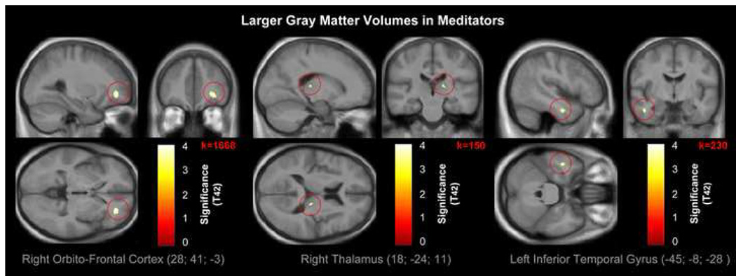
Figure 1. Larger GM volumes in meditators.
Views of the right orbito-frontal cortex (left panel; p<0.04FEW-corr), right thalamus (middle panel; p<0.0005uncorr), and left inferior temporal gyrus (right panel; p<0.0005uncorr), where GM is larger in meditators compared to controls. The color intensity represents T-statistic values at the voxel level. The results are visualized on the mean image derived from the 44 T1-weighted scans of the subjects analyzed, and presented in neurological convention (right is right).

