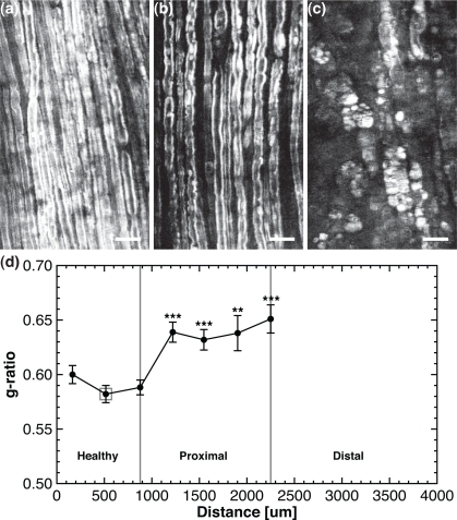
Fig. 4.
Histomorphometry on ex vivo crushed sciatic nerve 1 week post-injury. (a)–(c) Snapshots of the healthy, proximal and distal region of the lesion respectively, corresponding to the 3 ROIs of Fig. 3. (d) The g-ratio versus the position along the crushed sciatic nerve. All scale bars are 25 μm.

